A Compressive Review on Newcastle Disease Virus in Ethiopia
Tel: 251911000000, Email: mtesfa6@gmail.com
Abstract
Newcastle disease is an acute viral disease that affects both domestic and wild bird species around the world. It is a costly and widespread disease in most poultry-producing countries, including Ethiopia. Newcastle disease virus is a member of the genus orthoavulavirus, species avian orthoavulavirus (AOAV-1), and a new subfamily Avulavirinae of the family Paramyxoviridae that causes Newcastle disease. The virus is a non-segmented, single-stranded, enveloped, negative-sense RNA virus. The virus's genome contains six open reading frames that encode six structural proteins and two non-structural proteins. The virus's two major surface glycoproteins are hemagglutinin-neuraminidase and fusion protein. The hemagglutinin-neuraminidase protein mediates virus binding to host target cells, whereas the fusion protein facilitates viral envelope fusion with the cellular membrane of the target cells. Based on serological and phylogenic examination of the virus, fifteen distinct avian orthoavula virus serotypes are present. Strains are further grouped into three main pathotypes depending on their virulence and clinical signs: velogenic, mesogenic, and lentogenic. Newcastle disease is the most challenging avian disease in Ethiopia, resulting in significant economic losses for the poultry sector. The disease causes abrupt death with a 100% fatality rate to subclinical infection in chickens. The economic losses are associated with high mortality, morbidity, disease containment measures, outbreak eradication, and decreased egg production from breeder flocks. Regular outbreaks of the disease have posed a severe threat and confronted Ethiopia's burgeoning chicken sector. In endemic nations, there is a need to strengthen prevention and control strategies for Newcastle disease. As a result, this review article provides current scientific information on the Newcastle disease virus, including pathogenesis, antigenic variants, genetic diversity, current taxonomic classification, epidemiology, and various diagnostic techniques, in order to highlight the disease's control and prevention directions. Its goal is to combine numerous study findings from various sites and assess the disease's state in Ethiopia. Finally, to emphasize the poultry industry's economic importance in the country and to make recommendations for efficient management and prevention measures.
Keywords
Newcastle disease virus • Endemic • Genetic diversity • Diagnostic technique
Abbreviations
AAV: Avian Avulavirus; AHA: Animal Health Australia; AIV: Avian; Influenza Virus; AOAV: Avian Orthoavulavirus; APMV: Avian Paramyxovirus; CDNA: Complementary Deoxynucleotide Acid; CEF: Chicken Embryo Fibroblast; CPE: Cytopathic Effect; CSA: Central Statistical Agency; DF-1: Immortal Chicken Embryo Fibroblast Cell; EDTA: Ethylenediamine Tetra Acetic Acid; ELISA: Enzyme Linked Immunosorbet Assay; F: Fusion Protein; FAO: Food and Agricultural Organization; HA: Hemagglutination; HAU: Hemagglutination Unit; HI: Hemagglutination Inhibition; HN: Hemagglutinin-Neuraminidase; ICPI: Intracerebral Pathogenicity Index; IVPI: Intravenous Pathogenicity; MDT: Mean Death Time; MRNA: Messenger Ribonucleic Acid; ND: Newcastle Disease; NDV: Newcastle Disease Virus; OIE: International Animal Health Organization; PBS: Phosphate Buffer Saline; RBC: Red Blood Cells; RNA: Ribonucleic Acid; RNP: Ribonucleoprotein; RPM: Revolution Per Minute; RT-PCR: Reverse Transcription Polymerase Chain Reaction; SPF: Specific Pathogen Free; VNT: Virus Neutralization Test; VTM: Viral Transport Media
Introduction
Poultry farming is a segment of the livestock sector that is involved in considerable agricultural activity in practically every developing community, mostly in Africa. It is one of the fastest-growing segments of global agricultural demand since it provides a unique benefit to the sector while also raising community living standards [1]. This is due to its quick yield return, low investment requirements, short generation intervals, and quick reproduction cycle when compared to most other livestock [2]. It’s also one way to get food and ensuring food security in underdeveloped nations such as Ethiopia, where poverty and undernutrition are prevalent [3].
Ethiopia boasts Africa's greatest livestock population. The country's current total poultry population is 48.96 million [4]. Ethiopia has a population of 40,001,033 indigenous chickens (81.7%). The country's estimated exotic and hybrid poultry populations are 3,637,250.00 (7.43%) and 5,317, 392.00 (10.86%) million, respectively. Both exotic and indigenous poultry contribute much less to Ethiopia's economy than in other African countries. Disease issues, inadequate management, and breeds with low genetic potential hinder economic improvements in most parts of the country. The main causes of eviction are outbreaks of ND and infectious bursal disease [5].
Newcastle Disease Virus (NDV) is a member of the genus Orthoavulavirus and the species Avian Orthoavulavirus (AOAV-1) [9]. The virus is a single-stranded, non-segmented, negative-sense RNA virus with a helical shape and a molecular weight of 5-5.4x10⁶ daltons. The virus's genome comprises six open reading frames, each of which encodes six key structural proteins. Hemagglutinin-Neuraminidase (HN), Large Polymerase (L), Phosphoprotein (P), Matrix (M), Fusion (F), Hemagglutinin-Neuraminidase (HN), and Nucleoprotein (N). The virus's two major surface glycoproteins are the HN and F proteins. The HN protein aids in the virus's binding to host target cells, while the F protein aids in the fusion of the viral envelope with the target cells' cellular membrane [10].
Based on serological and phylogenic examination of the virus, fifteen (15) distinct avian orthoavula virus serotypes identified [11]. One of the most limiting characteristics of distinct strains of the virus is the presence of diversity in pathogenicity for chickens. Based on their virulence and clinical symptoms, strains are further divided into three pathotypes: velogenic, mesogenic, and lentogenic [12]. Velogenic strains are also classified into neurotropic or viscerotropic depending on their predilection site following infection of the chickens and associated with high mortality [13].
Infected hosts with ND experience respiratory, neurological, gastrointestinal, and reproductive issues. Exhaled air from diseased birds and contaminated food are sources of infection. Gasping, coughing, paralysis (wings and legs), and twisted necks are some of the most common signs of the condition [14]. Virus isolation from SPF embryonated chicken eggs or cell culture is the gold standard diagnostic procedure, followed by hemagglutination testing, hemagglutination inhibition testing, and virus pathotyping [15].
In 1926, the first cases of the disease were found in Newcastle-upon-Tyne. The name ND is given concerning the place it is found, and its name is coined by Doyle to avoid confusion with other diseases. Since then the disease is distributed throughout the world. In Ethiopia, the first confirmed case of the disease was recorded in 1971, following the occurrence of an outbreak in Asmara, Eritrea. The disease is then gradually spread across the country by wild birds and other risk issues. It has grown endemic in the village and commercial poultry populations, and it recurs every year, resulting in significant financial losses [16].
ND is the main challenging avian infection in Africa including Ethiopia since it is prevalent both in commercial and backyard chickens. Therefore, it is the most significant impediment to the development, survival, and productivity of Ethiopian chicken flocks, resulting in significant economic losses for chicken production.
The economic losses are associated with high mortality, morbidity, disease containment measures, outbreak eradication, trade restrictions, decrease egg production, and low-quality egg production from breeder flocks [17]. Virulent strains of the virus cause up to 100% mortality in unvaccinated chickens. Outbreaks of ND are limiting factors for poultry production in developing countries [18]. Regular outbreaks of the disease have posed a severe danger to Ethiopia's burgeoning chicken sector: As a result, the aim of this review manuscript is
• To provide up-to-date scientific information on NDV, including pathogenesis, genetic diversity, antigenic variants, epidemiology, and diagnostic approaches;
• To highlight the disease's combating direction;
• To compile research findings and the disease's status in Ethiopia; and to emphasize the disease's economic significance in Ethiopia's young poultry sector.
Literature Review
Definition
The Avain orthoavulavirusserotype1 is the etiological agent of ND affecting birds that meet one of the following virulent criteria. The virulent strains of NDV are defined by OIE that have an ICPI of greater than or equal to 0.7 in day-old chicks or a fusion cleavage site with multiple basic amino acids and phenylalanine at position 117. When three arginines or lysines are present between residues 113 and 116, it is referred to as multiple basic amino acids. A total of 109 countries had reported to the OIE in the last five years among 200 member countries since ND has a global impact. The virulent strain of the virus is one of the most overwhelming diseases of the poultry industry since it can cause 100% mortality in the susceptible chicken population [19].
History and distribution of the virus
Newcastle disease is a viral infection that affects avian hosts [20]. The first cases of the disease were found in 1926. In 1926, the first cases of the disease were found in Newcastle-upon-Tyne. The disease was named pneumoencephalaitis for the first time. Later on, the name ND is given where the place it is found, and its name is coined by Doyle to avoid confusion with other diseases. Doyle was the first person that discovered NDVvia laboratory experiment and noted it as not related to the Avian Influenza Virus (AIV). Since then ND is disseminated throughout the world and now has been known in most of the main poultry producing countries. The greater spread of the virus is linked to the commercialization of chicken and their feed. The spread of disease into the United States was aided by the transportation of caged birds, with psittacine and mynah birds being linked to disease epidemics.
ND, locally known as fengle, was initial found in 1971 in Asmera, and followed by spread to the country along transport routes. Outbreaks of the disease were recorded in Addis Abab in 1972, Harer, Shola, and Bishoftu (formerly Debre Zeit) poultry farms in 1974 NVI, 1974 In Ethiopia, ND is the biggest barrier to poultry farming. Velogenic strains of the virus are involved in the early outbreaks that can be caused up to 100% mortality. The disease is then gradually disseminated throughout the country via wild birds and other risk factors identified from various geographic locations. It has grown endemic in the village and commercial poultry populations, and it persists each year, resulting in significant financial failures. Prevalence rate of ND in commercial and village chickens is 80% and 43.7%, respectively.
Etiology and taxonomic classification
Newcastle disease is caused by the avian orthoavulavirus serotype 1. A uniform naming and classification scheme of viral isolates into evolutionary related groups has been predicted based on the whole nucleotide sequence of the fusion gene, phylogenic topology, and evolutionary distance. The evolutionary distance at the nucleotide level is used to assign new genotypes and subgenotypes. The species Avian Orthoavulavirus (AOAV-1) belongs to the genus Orthoavulavirus (OAV-1) in the Paramyxoviridae family's new subfamily Avulavirinae. The subfamily Avulavirinae is classified into three new genera: Orthoavulavirus, Metaavulavirus, and Paraavulavirus (ICTV, 2019).
Currently, fifteen (15) distinct AOAV 1 (AOAV-1 to AOAV-15) serotypes are found in several species of domestic and wild birds depending on the serological and phylogenic analysis of the virus. Serotype 1 remains an economically significant pathogen within the poultry industry. AOAV-2, AOAV-3, AOAV-6, and AOAV-7, on the other hand, have been documented to infect poultry. All the virus isolates belong to serotype 1 (AOAV-1). Based on their virulence and clinical symptoms, viral strains are further classified as velogenic, mesogenic, and lentogenic, which indicate high, moderate, and low virulence, respectively. The lentogenic strains are avirulent, causing very minor respiratory symptoms or no clinical indications in young chicks. In the business chicken sector, lentogenic strains of the virus are widely employed as live attenuated vaccines. The mesogenic strain shows nervous and/or respiratory signs with a low mortality rate compared to velogenic variants of the virus. The mortality incidence of ND is linked to the age of susceptible chickens. Juvenile chickens are more at risk than adult hosts. It also reduces egg production in laying flocks. Velogenic strains cause severe necrotic and hemorrhage in the gastrointestinal system and lymphoid organs. It can also produce nervous and respiratory symptoms, as well as a high likelihood of mortality at any age.. Lymphocytic tissues and the neurology system are prominent targets for the strains. Velogenic strains are the most acute and devastating strain of chickens, with unimmunized groups experiencing 100% mortality.
Results and Discussion
Genotypic characteristics
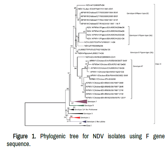
ND virus is a major threat to the fowl business globally since novel wild-type genotypes are explored each time. Different types of genotypes mingle in various fractions of the globe and continuously evolving. There are 55 full genomes of several NDV strains, with three different genome lengths: 1586 nt, 1592 nt, and 1598 nt. The extensive circulation of the virusin fowl flock showed to the major hereditary variety of the virus and the regular appearance of strains in the world. Two separate classes of NDV isolates (class I and II) can be found in a single genotype depending on genome length and nucleotide sequences. Class I viruses belong to a single genotype with three subgenotypes, whereas class II viruses are currently divided into 20 genotypes on the basis of comprehensive sequencing of the coding area of the fusion gene. Class II contains both vaccine and virulent viruses which are found in chicken and feral birds. The majority of the viruses found in class II groups are virulent and also highly diverse that continuously evolving. These viruses are responsible for considerable financial beating in the chicken industry around the world. Class I contains lentogenic strains identified from waterfowls birds, wild birds, and household chicken. The strain is less diverse and belongs to a single genotype that mostly avirulent to chickens. It has the longest genome sizes (15,198 nt) among AOAV-1 which is dispersed universally in feral birds and regularly identified from market samples. group I strains have the potential to evolve into virulent strains following circulation in chickens after acquiring mutations in the F and HN proteins. Therefore, the isolation and pathotype identification of class I strain in chickens facilitate monitoring the evolution of the virus. The sequence of the F gene protein is the basis for phylogenetic analysis of the virus and helps to evaluate the origin and distribution of disease outbreaks (Figure 1).
Genotype I, II, III, and IV was identified between 1930 s and 1960 s and are recognized as ancient genotypes. These genotypes have 15,186 nucleotides of genomic size. Class II genotypes that are currently evolving were investigated following 1960 s. There are at least twenty distinct genotypes identified depending on whole fusion gene coding sequence of class II NDV isolates. Three subgenotypes are present within genotype I of class I isolates with new nomenclature. These are 1.1.1, 1.1.2 and 1.2 with their respective former name of 1a, 1b and 1c and 1d. XIX, XX and XXI newly identified genotypes of class II viruses that are evolved from former genotypes and subgenotypes. XX and XXI two new genotypes emerged from genotype VI. Sub genotype Va has been modified into genotype XIX in the current classification system. All genotype XX viruses were isolated from hens in Asia and European countries between 1985 and 2011. Chickens and pigeons are a source of genotype XXI viruses in diverse Asian, European and African countries between 2005 and 2016. Genotype XXI virus is a clade of fowl isolated in Ethiopia which was identified as separate subgenotypes VIi, VIf and VIh. According to current updated classification and nomenclature systems, the former subgenotypes VIf and VIh are now known as VI.1.2.2.1 and VI.1.2.1.2, respectively. Genotype II and VII (VIId) are also found in Ethiopia depending on sequencing of the F gene coding region. The most diverse genotype among all NDV genotypes is genotype VI which can be isolated in all continents except Antartica. Former sub genotypes VII have been amalgamated into three sub genotypes with their new nomenclature depending on current classification system set by. These are VII.1.1, VII.1.2 and VII.2. Sub genotype VII.1.1 and VII.2 are responsible for the fourth and fifth panzootics respectively in different countries.
Morphology and genomic organization
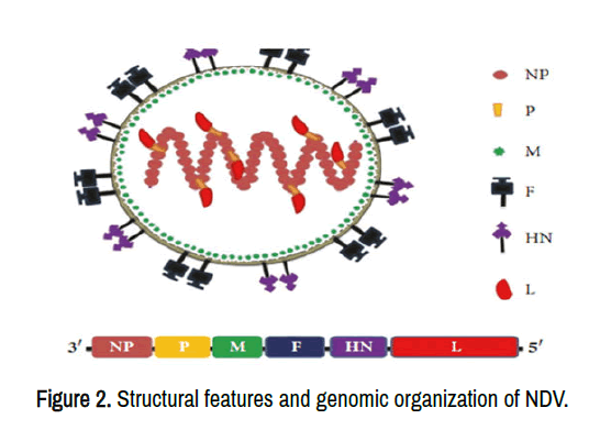
The virus has pleomorphic structures based on electron microscopic examination of the virus with diameters of 300 to 500 nm. It is a lipid enveloped particle derived from the host cell membrane with the projection of F and HN spear conjugated protein (Figure 1) that plays a role in the virus's infectious cycle.
The virus genetic material is nonsegmented, single-strand, negative-sense RNA genomes with a 15 kb genome containing six open reading frames that code for six structural and two nonstructural peptides: Nucleoprotein (NP), Phosphoprotein (P), Matrix protein (M), fusion protein (F), Haemagglutinin-Neuraminidase (HN) protein, and RNA directed RNA polymerase (L). 3'-NP-P-M-F-HN-L-5 is the linear order of the genes (Figure 2). RNA editing during P gene transcription produces the non-structural proteins V and W. The transcriptional control of each gene is aided by short coding sections present between each gene. The leader and trailer sections are found at the 3' and 5' ends of the genome. The brief extragenic leader and trailer sequences flank each gene. Gene boundaries are separated by Intergenic Sequences (IGS). The trailer sequence is 114 nucleotides stretched, while the leader sequence is 55 nucleotides. The transcription, replication, and packaging of genomic and antigenomic RNA are all regulated by the leader and trailer regions. Transcriptional regulatory sequences are preserved at the start and end of each gene. Multiple basic amino acids, as well as phenylalanine (F) at position 117, are found in virulent variants of NDV's fusion cleavage site.
HN and F proteins are important exterior glycoproteins involved in virus infection, tropism, pathogenicity, and antigenicity. Altering the F protein's glycosylation location boosts the virus's virulence and pathogenicity. The HN protein facilitates the fusing of the viral envelope with the cellular membrane of the target cells to commence virus infection, while the F protein facilitates the binding of the virus to host target cells. The viral envelope is made up of the M, HN, and F proteins. The matrix protein is situated beneath the envelope and plays an important function in the virus's development. NP, which is firmly linked to the genomic RNA, forms the virus's core structure. NP, P, L proteins and RNA genome forms the Ribonucleoprotein complex (RNP) whereas the spike-like protrusions are made up of HN and F proteins. These surface glycoproteins are the main protective antigen that induces neutralizing antibodies during infection. The F protein is the main antigenic determinant compared to HN proteins. RNP is a template for RNA transcription and replication.
Biological properties
Haemagglutinin-Neuraminidase protein (HN): HN protein is a multifunctional surface glycoprotein with a cytoplasmic domain, a transmembrane region, a stalk region, and a globular head that contains the receptor binding site as well as all antigenic sites. The receptor-binding site is accountable for the affection of the virus to its sialic acid-containing receptors. It also has haemagglutinating and neuraminidase activity, which it uses to hydrolyze sialic acid molecules from offspring viral particles in order to prevent viral self-aggregation. Neuraminidase hydrolyzes the ketosidic bond between substituted neuramic acids on host receptors, allowing the F protein to make contact with the host membrane and the virus to enter the cell. That globular head contains the main amino acid residues implicated in sialic acid receptor binding and NA activity. HN protein has seven antigenic sites that overlap. The only linear epitopes found in the HN gene are residues 345 to 353; these are vulnerable to immunological pressure, resulting in antigenic variation. The antigenic disparity would be bolstered by differences in the linear epitopes of the HN protein. Through its association with the Fusion protein, the HN protein promotes membrane fusion, allowing viral RNA to enter the host cell.
The pathogenicity of the virus in chickens is determined by the length of the HN protein. Depending on the strain, the length spans from 571 to 616 amino acids extended open reading frames of 6 to 45 amino acids are found in longer HN genes. The virus is considered a velogenic strain when HN gene has 571 amino acid lengths while virulent strains of the virus have 616 amino acid long. Though HN length of 577 amino acid can be found in multiple pathotypes of the virus. The involvement of virulence with the length of the HN gene is due to the requirement for the cleavage of HN0 precursor in avirulent viruses to become biologically active. The nucleotide and amino acid sequences of the HN protein reveal the virus's strain-dependent structural variations.
Fusion (F) protein: The protein facilitates viral entry into the host cell by fusing the viral envelope with the plasma membrane of the host cell at neutral pH. F protein is the most important determinant of pathogenicity. The F and HN proteins are defensive antigens that stimulate virus-neutralizing antibodies during viral infection by forming spike-like projections on the outer surface of the viral membrane. The host cell protease enzyme cleaved inactive precursor (F0) into physiologically active F1 and F2 subunits for virus entry and cell to cell interaction which results in the initiation of infection. Disulfide linkages connect the F1 and F2 subunits. The cleavage site sequence of the F protein is a key pathogenicity determinant in chickens. The F gene is used to determine phylogenic analysis and evolutionary distance. The F gene is usually selected for comparative analysis since it shows genetic distinction than other internal nucleocapsid genes.
Lentogenic, mesogenic, and velogenic strains have distinct amino acid sequences at the F cleavage site. Extracellular proteases confined to certain tissues break the monobasic cleavage site found in all lentogenic stains. The multibasic cleavage site is cleaved by intracellular protease enzymes in all mesogenic and velogenic strains. Therefore, the F gene is used to distinguish lentogenic strains from mesogenic and velogenic strains depending on sequence changes at the fusion cleavage spot.
Nucleoprotein (NP): Nucleoprotein is the structural subunit of nucleocapsid. The virus's nucleocapsid configuration is formed by the most copious protein. During genome expression, the protein is connected with the viral Polymerase (P-L), and during nucleocapsid assemblage, it is coupled with the P protein. It is the viral genome's primary regulator in terms of replication and transcription and, highly immunogenic since it induces antibody response in chickens. The amino acid series inferred from the NP gene nucleotide chain reveals an equal number of positive and negative charges. The NP molecule interacts with the negatively charged phosphoprotein, RNA backbone, and positively charged membrane or matrix protein during virus replication.
Large polymerase protein (L): The L protein, which is linked to the NP and P proteins, is an RNA-dependent RNA polymerase. It is the major viral protein that has all agitator actions and catalyzes the replication and transcription of viral mRNA. And it involves synthesizing the complementary stand, methylation, capping, and polyadenylation processes of newly synthesized RNA. The viral polymerase binds to the genomic RNA at the 3 leader promoter and then transcribes all viral mRNA with decreasing efficiency. The viral genome replication is reliant on the intracellular concentration of N protein. The polymerase enzyme copies the entire genome devoid of recognizing transcriptional signals, resulting in a genome that is neither capped nor polyadenylated.
Matrix protein (M): Hydrophobic protein with many basic residues, and maintain the shape of the virus. Early in infection, the protein is found in the nucleus and then penetrates the cytoplasm, where it adheres to the cellular membrane, allowing for later infection. The protein interposes between viral envelope and nucleocapsid. It is functioning for the packaging, budding and release of newly assembled viral particles in the protoplasm and the plasma membrane by interacting with nucleocapsid, lipid bilayer, and surface glycoproteins. It controls how viral copying and transcription are balanced. The M protein helps to assemble the maturation and infection processes in time and space, contributing to the sequence of events during budding and fusion. Matrix peptide is the main conserved amino acid among the paramyxoviruses that helps to establish a consistent classification of the viruses. The amino acid sequence of the M protein is extremely homologous on behalf of different strains of the virus.
Molecular and biological basis of virulence determinants
Pathogenicity is the capability of the organisms to induce infection in the host. Virulence is the extent of pathogenicity and more quantitative. Biological (in vivo) and molecular (in vitro) experimental testing can both be used to determine the virus's pathogenicity. The most significant pathogenic marker of the virusis found in F protein. Major factors of virus pathogenicity include the amino acid sequence at the F cleavage site and the capacity of distinct host cellular protease enzymes to cleave different pathotypes of proteins. To make the progeny virus infective, host cell proteases should split the precursor Fusion protein (F0) into F1 and F2 proteins. A disulfide bond connects these two proteins, which may aid virus adhesion to the host plasma membrane. The C-terminus of the F2 protein and the N-terminus of the F1 protein of lentogenic strains have a monobasic amino acid motif. The lentogenic amino acid motif was broken by an extracellular protease enzyme identified in the respiratory and intestinal system. Multiple F cleavage sites at the C-terminus of F2 protein and phenylalanine(117) at the N-terminus of F1 protein are present in mesogenic and velogenic strains, and these sites are cleaved by intracellular furin-like protease enzymes. The amino acid sequence of velogenic and mesogenic strains of the viruses are 112R/K-R-Q-K/R-R-F117 at the C-terminus of the F2 proteins and the N-terminus of F1 protein. Lentogenic strains of the virus have 112G/E-K/R-Q-G/E-R-L 117 amino acid sequences. Except for pigeon paramyxovirus 1, all virulent ND viruses possessed a critical amino acid at position 112.
In vivo experimental testing can also be used to determine the pathogenicity of strains (Table 1). In-vivo pathogenicity of the virus has been defined and quantified using Mean Death Time (MDT), Intracerebral Pathogenicity Index (ICPI), and intravenous pathogenicity index. The ICPI is the method of choice for determining NDV pathogenicity. However, the entire procedures are time-consuming that takes at least five days. Therefore, rapid procedures for distinguishing pathotypes of virus strains and isolates are necessary. The sequence analysis, restriction enzyme digestion, and molecular hybridization of the F gene fragment, as well as one-step RT-PCR, are faster diagnostic techniques for distinguishing strain pathotypes.
| Pathotypes | MDT (hr) | ICPI | IVPI |
|---|---|---|---|
| Velogenic | <60 | >1.5 | 2 |
| Mesogenic | 60-90 | 0.7-1.5 | 0-0.5 |
| Lentogenic | >90 | <0.7 | 0 |
MDT= Mean Death Time, measured in hours to death; ICPI= Intracerebral Pathogenicity Index, based on an average score of clinical signs over time (min=0.00.max=2.0)
Epidemiology
ND is found all over the world and OIE notifiable disease. Epidemiological data revolving in chick and undomesticated bird flocks within a detailed province can give practical information for ND management and deterrence. Four major economically overwhelming panzootic have occurred in the last nine decades. The first panzootic have been started in Asia and Europe in the mid-1920 s. The disease took around 20 years to spread throughout the rest of the planet. The second panzootic has become fully distributed within four years most likely due to the increased commercialization of the poultry sector and increased global trade of imprisoned pen birds across the world. Genotype VI isolates in racing pigeons was assumed to be the origin of the third panzootic. A number of species of birds were affected and has become difficult to control the disease since devoid of absolute control mechanisms of racing pigeon’s husbandry. Finally, the fourth pandemic, which began in the 1980 s and has been related to numerous economic losses in a variety of countries, is now underway. Genotype VII groups of the virus cause the occurrence of the fourth pandemics and the most rapidly evolving varieties of the virus currently.
In Central and South America, Asia, and Africa, the disease has spread to epidemic proportions. It first appeared in Europe on an irregular basis in 1981, and then quickly spread over the globe. It is endemic to Southeast Asia, some North and South American countries, and Africa, where it causes significant economic losses in commercial chicken farms. All poultry species are affected by ND, but the pathogenicity differs from the host. Virulent strains of the virus commonly infect cormorants, pigeons, and psittacine species and served as a source of infection for poultry. Low virulent strains are widespread in chickens and waterfowl birds. Low virulent strains are prone to lower productivity in fowls.
Host range: The virus can infect a wide spectrum of hosts. It has the potential to impact over 241 domestic and wild bird species from 27 of the 50 bird orders. NDV can infect a variety of bird species spontaneously or in a lab setting. The disease mainly affects chickens although all species of birds are most likely susceptible to the disease. The susceptibility of the host might diverge significantly among avian species based on varieties of the virus. Virulent strains of the virus can be isolated from all types of village and commercially reared chickens. Chickens are thought to be the virus's most vulnerable and usual host. Virulent strains cause serious disease in chickens with 100% mortality among domestic poultry. In general, chickens, turkeys, and pigeons are the majority vulnerable species of birds while ducks and geese are the slightest at risk birds. Ducks may not show clinical signs. All age groups of the birds are susceptible to the disease. However, the severity of sickness differs between juvenile and adult birds. For the time being, anyone who come into intimate contact with the virus may acquire a localized eye infection (conjunctivitis). The virus is also frequently identified from untamed migratory marine birds which are less virulence for chickens. Several outbreaks during the last two or three decades have implicated wild birds, captive birds, and racing pigeons in the introduction of virulent strains of the virus to poultry. Psittacine birds excrete virulent strains intermittently for long periods with no presenting clinical signs that play in dissemination of the virus.
Source of infection and transmission: ND is an extremely contagious and devastating viral infection that can infect a wide range of domestic and wild bird species around the world. The disease has manifold possible ways of dissemination. The capability of viral agents to remain in the environment has the potential to assist disease transmission between species. The disease spread mainly via direct and indirect contact between healthy and infected birds. The ND virus is transmitted and propagated via virus-containing feces and discharge from affected hens. The virus's excretion route is determined by the organs where it replicates and differs depending on the strain. Aerosol transmission is the major means of ND infection in intensive production systems. In village chickens, primary way of transmission is through route. The disease is spread from one bird to the next and from one farm to the next primarily through aerosol, but it can also be spread through contaminated feed, water, feces, eggs, and the carcass of sick birds. Natural routes of infection such as oral, nasal, and ocular appear to give emphasis to the disease's respiratory natureDuring the incubation period, infected birds begin to excrete viruses, which are then shed in aerosol, respiratory discharges, and feces. The incubation period of the virus is 2-15 days post-infection of the host. The virus is secreted for up to 1-2 weeks following infection in chickens, turkeys, grouse, and pheasants. Psittacine Birds that are parrots, parakeets, and macaws can excrete infectious viruses for a number of months to one year after infection.
Risk factors and physicochemical nature of the virus: A range of risk factors is present for the incidence of ND in avian hosts. The risk factors that boost the frequency of the infection are geographical location of the farm, distance between neighboring poultry farms, age of the host, and structure of the flock, restocking chickens from markets, presence of migratory wild birds, poor biosecurity measures, insufficient sanitary, poor quarantine measures and inadequate vaccination programs appeared to have a significant responsibility in severity of disease. The occurrence of ND is different among years in Ethiopia. The persistence of the virus in the environment is awfully variable since it can be exaggerated by different issues like temperature, humidity, the suspending agent, and exposure to light. The virus is heat steady and remains infectious in the bone marrow and muscle of slaughtered chickens for six months at-20oC. These factors have all had a role in the disease's spread and perseverance on farms. The other significant feature of the virus is its susceptibility to disinfectants containing detergents since the lipid layer present in the envelope of the virus. Dyes, lipid solvents, formaldehyde, and oxidizing substances are all toxic to the virus.
Antigenic and genetic diversity
There is genetic and antigenic diversity of AOAV-1 strains circulating depending on genetic characterization of the F gene although all isolates of the virus belonging to serotype one. The antigenic and genetic assortment is recognized within the AOAV-1 serotype. The extensive circulation of thevirus in chicken flocks led to the major hereditary variety of the virus and the regular appearance of strains in the world. The constant multiplication, movement, and preservation of lentogenic strains among avian host poses a significant risk of virulent variants emerging. The evolutionary dynamics of this RNA virus are mostly influenced by different sorts of mutations, as they are for other RNA viruses. This evolutionary changes/modifications of the virus are determined by polymerase error, genetic recombination, antigenic drift and shift, natural transmission between different hosts, negative and positive selection pressure of the fusion protein cleavage site. The evolutionary rate, selection pressure, and phylogenesis all have a role in the host shift and virulence change.
The fast mutational rate of ND viruses results in a high genetic variety/antigenic subtypes of the viruses, as well as the frequent emergence of more virulent variants that can overcome acquired immunity and survive in the population. A plausible source of velogenic strains could be mutations of lentogenic strains of the virus kept in chicken and wild bird reservoir hosts. A number of lineages or genotypes have been recognized among the AOAV-1 since the appearance of NDV in 1926. Different genetic groups of the virus go through evolutionary changes at the same time in diverse parts of the planet. The genetic basis of antigenic variation is the sequencing of the F gene of multiple different strains and the identification of escape mutants. Antigenic variation is one of the most efficient ways for a virus to evade neutralizing antibodies and elude the host's immune system. Neutralizing epitope variants emerged following point mutation in the F gene.
The disease's control, prevention, and diagnosis are all made more complex as a result of this evolutionary transition. The failure of standard diagnostic techniques to detect novel genetic variations of the virus has also been linked to evolutionary changes in the genome. These lead to significant economic losses in poultry industries. Therefore, knowing the genetic variation and detecting newly circulating variant viruses provides important approaching into the virus's methods for using altered structure and makes future vaccine creation easier. Besides, these also have significant importance for the development of new diagnostic tests for the control and prevention of the disease.
Pathogenesis
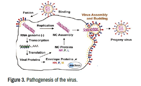
A complex interaction between the virus and the infected hosts is the pathogenesis of the virus. Infected hosts experience harm as a result of the encounter, which leads to mortality or death. The virus infects the host cell once it recognizes its receptors and binds to sialyglycoconjugates on the cell surface. This is followed by the fusion of the viral envelope with the host cell membrane which is facilitated by viral surface glycoprotein interactions, HN and F. Then viral genome is released into the cytoplasm. Replication, transcription, and translation take place in the cytoplasm of the host cells, whereas assembling or packaging of virus particles and the progeny mature in the plasma membrane via budding (Figure 3). ND virus replicates in different types of organs such liver, spleen, brain, and lymphoid tissues. Finally, infection results and the appearance of different types of clinical features followed by the death of the infected hosts. Pathogens, host species, route of transmission, and environment are combinational factors for the pathogenicity of the virus. The strains of the infecting virus are the most important factors for the pathogenicity of the virus.
Virus replication
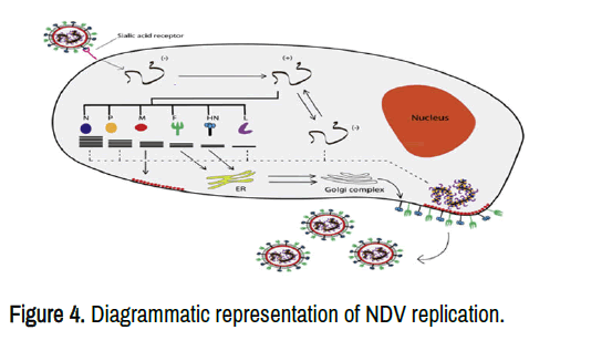
The virus infects a wide range of cells that contain sialic acid residues. Sialic acid is a receptor for the virus. The HN glycoprotein binds to sialic acid receptor found in the host cell, and a conformational change takes place both in HN and F protein. This leads to the experience of fusion peptides to the intent membrane that consents the fusion of the viral envelope and the surface membrane of red blood cells. A neutral pH has obligatory for fusion of paramyxovirus in the host plasma membrane. The low PH also mediated conformational changes of glycoproteins for induction of fusion or hemolysis of other enveloped paramyxovirus. Following fusion, the M protein dissociated from the nucleocapsid, and the viral genetic material is delivered to the cytoplasm to start replication and transcription processes (Figure 4). The active transcriptase complex contains NP, L, P, and genomic RNA. Full-length positive RNA is synthesized following genomic replication and serves as a template for the synthesis of negative genomic RNA. NDV replication adheres to the rule of six, which states that the length of the viral genome must be a multiple of six.
Transcription and translation
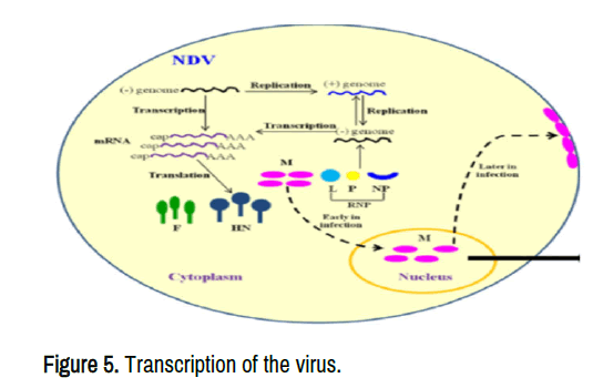
Transcription is the process by which the information in a strand of DNA is copied into new molecules of messenger RNA (mRNA). The transcription of NDV is comparable to that of other nonsegmented negative-sense RNA viruses. Following entry into the host cell cytoplasm, the viral genome has been transcribed and replicated. The negative-sense RNA genome is transcribed into positive-sense mRNA, which is then translated into viral proteins (Figure 5). Transcription begins with a 3' leader sequence and proceeds to the synthesis of mRNA from gene start to gene end sequences using viral RNA polymerase. The synthesis of the leader sequence stops at the first NP gene's border and is replaced by mRNA synthesis at the NP gene's gene start. Transcription continues in a start-stop mode until the mRNA of the last gene L is synthesized. The NP, P, and L proteins are necessary for nucleocapsid assembly. The assembly and bunding of mature viruses depend on the matrix protein and lipid raft over the cell membrane. Positive-sense RNA serves as a template for making negative-sense genomic RNA.
Clinical features and pathological lesion
The incubation period is the time interval between infection and illness development in the infected host which varies from 2-15 days depending on several factors. ND causes a wide spectrum of clinical diseases in avian species ranging from asymptomatic to 100% mortality depending on the viral pathotypes and species of hosts. The major clinical signs of the disease are gasping, coughing, sneezing, greenish diarrhea, corticoids, cyanosis of comb and wattle, tremors, paralysis (wings and legs), twisted neck. Chickens and turkeys are particularly vulnerable to velogenic NDV infection, but ducks and geese might not display any signs at all. Many types of psittacine birds and pigeons may carry vNDV devoid of showing signs. The severity of infection depends on virulence and tropism of the virus strains, host species, immune status, and route of infection, age, and environmental conditions. Intravenous inoculation causes neurologic symptoms, whereas aerosolization of a large viral dose affects the infected host's upper respiratory system.
Young hens dying from the peracute type of the disease may not have any visible signs of the sickness. Petechial hemorrhages and ulcers with elevated borders on the proventriculus mucosa, pneumonic lungs, and hemorrhages in the trachea, air sacs, brain, spleen, and other organs are the most usually detected gross postmortem lesions. Lesions in the gastrointestinal tract progressively become oedematous, hemorrhagic, necrotic, and finally ulcerative. Small, flat, red, or purple (petechial) hemorrhages may be seen on the breast muscle, heart muscle, and peritoneal adipose tissue and serosal surfaces.
Diagnostic techniques
History, clinical indicators, necropsy findings, serological, and molecular diagnostic approaches can all be used to diagnose ND. The disease clinically resembles highly pathogenic avian influenza virus. Therefore, accurate and rapid diagnosis is vital during outbreak occurrence to manage and avert distribution of the infection. Isolation of the virus is the golden diagnostic method. An enzyme-linked immunosorbent assay (ELISA), a viral neutralization test (VN), a hemagglutination inhibition (HI) test, a reverse transcription-polymerase chain reaction (RT-PCR), or a real-time RT-PCR test can all be used to provide a definitive diagnosis for NDV.
Clinical diagnosis: In both living and dead birds, the ND displays distinct characteristics symptoms and post-mortem lesions. The virus's primary diagnosis is based on the flock's history, clinical indicators, high mortality and postmortem lesion findings. A histological diagnosis of the disease is made by recognizing pathological lesions in several organs. In medical infections, clinical indications of infection emerge following an incubation period of 3-4 days on average. The major clinical signs of the disease are gasping, coughing, sneezing, greenish diarrhea, cyanosis of comb and wattle, paralysis (wings and legs), twisted necks. Petechial hemorrhages and ulcers with elevated borders on the proventriculus mucosa, pneumonic lungs, and hemorrhages in the trachea, air sacs, brain, spleen, and other organs are the most usually detected gross postmortem lesions.The disease is diagnosed in the lab by recognizing particular antibodies to the virus or by detecting the virus in tissues using immunological or molecular diagnostic techniques. The most conclusive diagnosis of NDV is the isolation and identification of the agent.
Histopathology: For a histopathological diagnosis, tissue specimens such as the spleen, proventriculus, duodenum, lung, trachea, and cecal tonsils are examined. The tissue specimens should then be desiccated in ethanol, set in paraffin, and sliced at a breadth of 4-6 um, after which they should be stained with Hematoxylin and Eosin for microscopic analysis. The presence of abnormalities in the lung, trachea, gut, proventriculus, spleen, and brain is used to make a histological diagnosis. This diagnostic technique can identify both acute and chronic manifestation of infection.
Pathogenicity index test: There is great disparity in pathogenicity of diverse NDV isolates with the type of avian species affected. Three internationally recognized in vivo assays are used to determine the pathogenicity of strainsMDT, ICPI, and IVPI have all been utilized in vivo tests to identify and enumerate the pathogenicity of the ND virus.
The average score per chicken per observation over 8 days is ICPI, which ranges from 0.0 to 2.0. The virulent strains provide scores of 2.0, while the lentogenic and asymptomatic enteric strains scores close to 0.0 and/or depending on the above scores, NDV can be pathotyped as lentogenic (<0.7), mesogenic (>0.7-1.3), or velogenic (>1.5).
MDT is done on ten SPF chicken embryos that are nine days old. A minimal lethal dosage is the greatest viral concentration that kills all embryos. If the inoculated SPF chicken embryo dies in less than 60 hrs, the virus is pathotyped as velogenic strains. Mesogenic strains when the embryos dead within the ranges of 60-90 hrs while lentogenic strains have more than 90 hrs. Intravenous Pathogenicity Index (IVPI) is the mean score per chicken per observation over 10 days in six old SPF chickens. Lentogenic and mesogenic strains have IVPI values of 0, while the virulent strains will approach 3.0.
Isolation of virus in embryonated chicken eggs: Isolation and cultivation of ND virus can be carried out in SPF embryonated chicken eggs and cell culture followed by HA and HI test. The virus might be demonstrated in a variety of organs and swabs in the acute stages of disease in the first 3 to 4 days subsequent demonstration of clinical signs. The areas of virus reproduction and viral shedding dictate the types of samples requisited for virus isolation. Embryonated hens' eggs are the most responsive and expedient vehicle for the proliferation of the virus and universally used diagnostic method.
Cell culture isolation: The virus could propagate in different types of tissue cultures of avian and non-avian species of source. Chicken embryo liver cells, chicken embryo kidney cells, chicken embryo fibroblast and African green monkey kidney cells are the most widely used avian and non-avian origin of cell lines. Avian origin primary cell cultures are highly susceptible to propagate NDV. The virus can be isolated using primary Chicken Embryo Fibroblast (CEF) or immortalized chicken embryo fibroblast cell cultures (DF-1). The growth of the virus is detected depending on the distribution of monolayer, level of cytopathic effect, formation of syncytia, and plaques in cell culture lines. CPE is characterized by granulation, vacuolization, clumping, syncytia formation of CEF/DF-1 cell monolayer due to infection by NDV. At the end of each passage in cell culture, the HI test should be performed again to determine the presence of the virus in the cell culture. Mg2+ ions and diethylaminoethyl dextran or trypsin are required for effective replication and plaques formation of lentogenic strains in the culture medium.
Serological diagnosis: The virus is investigated serologically using VN, ELISA, and HI tests. These tests are used to track immunological responses after vaccination and to identify flocks that are naturally sick. HI and ELISA commonly employed to detect antibodies in sera. In endemic areas, serological diagnosis receives little attention because assays cannot distinguish between antibodies generated by pathogenic viruses and those evoked by vaccines. Serology tests are also required to ensure that flocks are disease-free. Individual serum samples from the flocks under investigation should be taken in sufficient numbers.
Hemagglutination and haemagglutination inhibition test: Hemagglutination experiment determines the existence or absence of viral hemagglutinin in the allantoic fluids of eggs inoculated with the virus. It measures the quantity of hemagglutinin present. Hemagglutination is caused by the interaction of the HN protein with the receptor cell on red blood cells.
The HI test is detecting antibodies to the NDV. It is the most commonly used test that measures antibody titer and shows the accurate diagnostic result is the HI test. Positive specimens from hemagglutination can be utilized in an HI assay with NDV-specific serum to evaluate whether they are positive. NDV antibodies will bind with antigen and agglutinate RBC if the specimen has the virus. The quantity of virus in the antigen, fraction, and type of RBC, and the temperature at which the test is undertaken determine the outcomes of the HI test. Avian influenza virus, avian paramyxovirus serotypes, some adenovirus, and some bacteria can also agglutinate chicken red blood cells. Avian orthoavulavirus serotypes demonstrate a few levels of cross-reactivity among serotypes. There is cross-reactivity between AOAV-1 and AOAV-3 or AOAV-7. Monoclonal antibodies specific to AOAV-1, AOAV-3, and AOAV-7 can solve the cross-reactivity problems. The use of monoclonal antibodies also allows the characterization of antigenic distinctions in varieties of AOAV-1.
Enzyme-Linked Immunosorbent Assay (ELISA): ELISA kits are found to detect the presence of the virus antibodies. It is the most widely used diagnostic test for demonstrating and quantifying antibodies to assess vaccination efficacy, natural field exposure, and maternal antibody titer. The test is a low-cost, simple, and quick test that can be performed on a large number of samples at once and is computer-assisted. Direct, indirect, competitive and sandwich types of ELSA present with different levels of sensitivity and specificity.
Molecular identification technique: Conventional diagnostic processes such as serological assays, virus isolation, and pathotyping methods are time-consuming, expensive, labor-intensive, and unable to discriminate particularly virulent NDV strains from avirulent strain viruses due to the widespread use of live vaccinations. Therefore, rapid nucleic-based tests for detecting circulating viruses have been developed all around the world. Sensitive and specific diagnostic techniques such as RT-PCR, real-time RT-PCR, and sequencing are more effective than traditional diagnostic instruments in detecting and distinguishing strains. RT-PCR and real-time RT-PCR are the most extensively utilized molecular diagnostic techniques for detecting the NDV genome. In comparison to traditional diagnostic approaches, the tests are more sensitive, specific, and labor-saving.
Reverse Transcription-Polymerase Chain Reaction (RT-PCR): For in vitro nucleic acid amplification, RT-PCR uses RNA as a template. The usage of RT-PCR was made possible by the invention of the retroviral reverse transcriptase enzyme in the 1970 s. The RNA-dependent DNA polymerase reverse transcriptase catalyzes DNA synthesis using RNA as a template.
One of the most often utilized molecular diagnostic procedures for identifying viral genomes from representative specimens is RT-PCR. It is designed to detect and identify the pathotypes of the virus simultaneously by targeting the F gene portion encompassing the F cleavage site. For the characterization and identification of existing and emerging viral strains, RT-PCR in combination with Restriction Fragment Length Polymorphism (RFLP) is a very functional and rapid method. NDV isolates are divided into three groups based on their RFLP digestion patterns: lentogenic, mesogenic, and velogenic strains. It can also amplify DNA directly from specimens without propagating either in embryonated eggs or cell culture, and allowing for the sequencing of amplified DNA even if the virus is detectable in trace amounts.
The three primary processes in RT-PCR are extraction of nucleic acid, transcription of RNA into cDNA and amplification of cDNA by PCR. To target virus-specific nucleotide sequences, transcription and amplification stages require short sequence matching oligonucleotide primers. It can be done in a one-step or two-step procedure. The reverse transcription and amplification process is performed in a single tube in one step experiment. A two-step experiment involves reverse transcription and amplification in separate tubes.
Real-Time Reverse Transcription-Polymerase Chain Reaction (RRT-PCR): RT- PCR is a variation of the standard PCR for recognition and quantification of either DNA or RNA virus in specimens. When compared to standard diagnostic approaches, it is very sensitive, specific, and time-consuming. The number of copies of a particular gene sequence can be determined using sequence-specific primers. And able to monitor the progress of PCR reaction, measure the amount of amplicon at each cycle, increase the dynamic range of detection and amplification. It can also eliminate post-PCR manipulations. The amount of amplified product is quantified at each stage using fluorescent probes or fluorescent DNA binding dyes during the PCR cycle. The sensitivity and specificity of RRT-PCR are determined by the design and concentration of primers, probes, thermocycler temperature settings, type and concentration of enzymes, and the presence of PCR inhibitors.
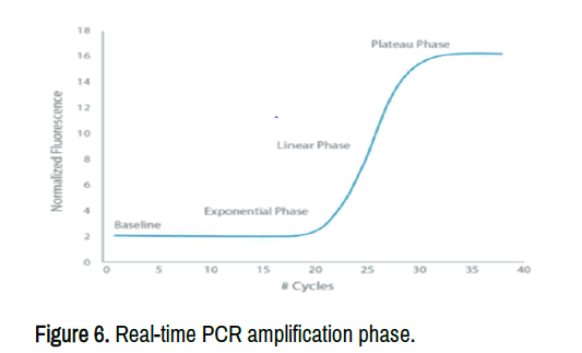
The real-time RT-PCR has three phases (Figure 6). Linear ground phase, exponential phase and plateau phase. PCR is just starting and the fluorescent signal is not above the background in the linear ground phase. The fluorescent signal is significantly increased and the optimal amplification stage has occurred with doubling PCR products in each cycle in the exponential phase. In the plateau phase, the substrate is worn out and Taq DNA polymerase is declining. The fluorescent signal will no longer increase at this stage. RRT-PCR can be done as a one-step or two-step procedure. In a one-step reaction, the entire reaction is carried out in a single tube, from cDNA synthesis to PCR amplification, whereas in a two-step reaction, reverse transcription and PCR amplification are carried out in separate tubes. One step RRT-PCR minimizes experimental variation and contamination since both enzymatic reactions are performed in a single tube. The starting template may prone to rapid degradation if not properly handled. It is less sensitive than two-step protocols. RRT-PCR is a novel and rapid practical instrument for the recognition of NDV.
ND virus RNA can be extracted from homogenized organs and swab samples or virus grown in SPF embryonated chicken eggs using commercially available RNA kits according to manufacturers' instructions. Then, at 37°C, incubate the mixture for 60 minutes. Finally, ethanol precipitation is used to extract nucleic acids, which are then resuspended in RNase-free distilled water or a suitable buffer. Until the RNA is processed, it should be maintained at -80°C. A spectrophotometer measures the purity and concentration of extracted RNA.
Prevention and control
There are no precise therapeutic agents for the ND. The virus's high level of interaction with physical agents is responsible for the virus's long-term survival in the environment. The virus' abolition from impacted countries appears to be impractical. Vaccines alone will not fix the problem unless they are used in conjunction with other techniques. Therefore, an effective immunization program, hygiene procedures, a reliable chick source, and quarantine and/or effective biosecurity practice are all essential components of a successful prevention and control program. Because the virus is discharged in vast volumes and resistant to external conditions, eradicating all sources of infection is nearly impossible. Cleaning and disinfection of dwellings between flocks, as well as all-in, all-out management, can help to lessen the virus's challenge. Halogen compounds and calcium hypochlorite powder, formaldehyde, iodophors, and oxidizing agents are the most successful antiseptics of the virus.
Vaccination
The precise strategy for preventing and controlling ND is determined by hygienic management, immunization schedule, degree and variety of maternally produced antibodies, and vaccine strain selection. Moreover, as several studies have shown, a consistent immunization program and stringent biosecurity measures are efficient approaches to control and prevent the disease. The vaccination programs are different depending on production systems, level of biosecurity and potential losses due to the disease. Breeders and layers are vaccinated against the disease, and oil-based vaccinations are used to provide long-term immunity before the start of egg production. Vaccines are widely used on commercial flocks, as opposed to extensive production systems. Inactivated and live vaccinations that generate protective immunity in chickens and other bird species are available all over the world. Although proper immunization protects chickens and other bird species from clinical sickness, the virus's replication and shedding persist, and it can infect neighboring flocks.
Due to their numerous advantages, live attenuated vaccines are the most commonly used vaccinations in chicken production. Lasota and Hb1 vaccines are the two most regularly used live vaccinations in various regions of the world. In many poultry production systems, ND4-HR and L2 are the two most commonly utilized live thermostable vaccinations. The vaccine can be produced in embryonated SPF eggs either from lentogenic or apathogenic strains of the virus. Apathogenic and lentogenic strains are economically feasible to use on a mass basis than an inactivated vaccines. Lasota is more invasive and immunogenic than Hb1 and boosts immunity effectively. The vaccines have been used to a greater extent than inactivated vaccines for many years. The route of vaccination depends on the age of chickens and the manufacturer's recommendation. Drinking water, aerosol, or ocular drops can all be used to give the live vaccination. Degree of attenuation, route of delivery, replication site, age and immune status of the chickens influence the immunity induced by the live vaccine. The main benefits of live vaccinations are that they are simple to use, relatively inexpensive, and provide reasonable immunity to both individuals and flocks.
An inactivated vaccine has no subsequent spread or adverse respiratory reactions since the agent has already been killed. The vaccine is significantly expensive than live vaccines. A large number of antigens are required for vaccination than live vaccines since the killed vaccine is not replicated in vaccinated hosts. The vaccine can be administered intramuscularly or subcutaneously. Of all these vaccine types, currently produced vaccines in NVI are LaSota, HB1 and Thermostable I-2 and commercially available for wider uses in large and small size commercial fowl farms in Ethiopia. Lasota and HB1 types of vaccines are class II genotype II viruses. Whereas I-2 type of vaccine is classified in class II genotype I virus originate from Australia
Public health importance
Newcastle disease can also infect humans besides avian species. Therefore, the virus is a human disease, and the most common symptom is conjunctivitis, which can develop within 24 hours of infection. For up to 7 days, the virus is shed in the ocular secretions. Conjunctivitis normally clears up after a few days. On rare cases, humans have developed a self-recovering influenza-like illness with fever and headache. Human-to-human transmission has not been proven, although human-to-bird transmission has.
Status of newcastle disease virus in ethiopia
In Ethiopia, ND is regarded as main serious chicken infection. In Ethiopia, the first acknowledged outbreak of ND was reported in 1971. ND continues to be a major limiting factor for both profitable and village poultry production systems, which are vital sources of income and food security for many countries, particularly in Africa. Detecting circulating genotypes is vital for investigating the molecular evolution and spread of the virus in the world. Therefore, understanding NDV assortment and dispersion is important for diagnosing disease patterns, evaluating spread by looking at links between isolates in different geographical locations, and predicting outbreaks. It's also crucial for keeping up to date on targeted biosecurity measures, vaccine investigate, and immunization methods. Genotype XXI virus is a clade of chicken isolated in Ethiopia which was identified as separate subgenotypes of VIi, VIf and VIh. According to current updated categorization and nomenclature systems, the former subgenotypes VIf and VIh are now known as VI.1.2.2.1 and VI.1.2.1.2, respectively, and have been linked to multiple outbreaks in the poultry production sector. Genotype II and VII (VIId) are also found in Ethiopia based on sequencing of the F gene coding region. The genetic divergence between Ethiopian genotypes found in the central and northern areas of the country is 25.1%. The Ethiopian velogenic strains viruses have 98-99% sequence identity at the nucleotide level with Sudan and Egypt isolates. Genotype VII plays a significant role in fatal infections in poultry and other susceptible birds.
The disease is widely spread throughout Ethiopia, yet the prevalence of the condition varies significantly, according to different research. A significant prevalence difference existed among breeds, season and geographical location. In comparison to indigenous hens, the disease's seroprevalence rate is higher in cross and foreign breeds. In intense manufacturing systems, the disease occurs more frequently than in extensive production systems. The poultry sector suffers considerable output losses as a result of ND on a national and worldwide scale. Millions of dollars are lost worldwide by ND. The disease is a well prevalent and endemic viral disease that affects chickens of all ages throughout Ethiopia. It has become serious challenge and danger to the poultry business in Ethiopia due to the emergence of antigenic variants of the virusas different studies indicated from different parts of the country.
The spread of the disease to native chickens is linked to the spread of improved chicken breeds from infected breeding and multiplication facilities. The key mechanism for the spread of disease across the country is the transportation of chickens from the countryside to towns for selling. The existence of the disease in Ethiopian village and commercial poultry chickens has been confirmed by serological and virological tests. In Ethiopia, the total seroprevalence of ND varies between 5.9% and 38.7% (Table 2).
| Study area | Seroprevalence (%) | Reference |
|---|---|---|
| Debre Berhan | 28.6 | (Tadesse, et al., 2005) |
| Rift valley and southern (Hawassa, Butajira, Alage, and Hossana) | 19.78 | (Zelek, et al., 2005) |
| Eastern Shewa zone | 5.90% | (Chaka, et al., 2012) |
| Central Ethiopia | 32.22% | (Tadesse, et al.,2005) |
| Bale zone (Sinana and Agarfa) | 27.86 | (Asfaw, et al., 2016) |
| Rift-Valley Areas (Debre zeit, Ziwaye and Tikur wuha) | 11.61% | (Terefe, et al., 2015) |
| Arsi zone | 38.70% | (Ambaw, 2014) |
| Alamat district | 26.20% | (Derbew, et al. 2016) |
| Nazareth | 38.33 | (Tadesse, et al.,2005) |
| Kersna Kondaltity | 5.6 | (Getachew, et al.2014) |
| Rift valley areas( Bishoftu, Batu, and Hawassa) | 12.7 | (Haile, 2017) |
| East Showa zone (Lume and Modjo districts) | 28.6 | (Jarso, 2015) |
| Central Ethiopia(Sebata Hawas district) | 11.34 | (Sori, et al., 2016) |
| North Gondar Zone (Dembia, Gondar city, Maksegnit and Amba Giorgis) | 34.60% | (Haile, et al., 2020) |
Conclusion
Newcastle disease is an acute and extremely infectious overwhelming viral disease that affects both domestic and wild species of birds across the world. The disease is widely spread throughout Ethiopia, yet the prevalence of the condition varies significantly, according to different research. The Avian orthoavulavirus serotype 1 is the etiological agent of ND. The species Avian orthoavulavirus (AOAV-1) belongs to the genus Orthoavulavirus 1 (OAV-1) in Paramyxoviridae family. Fifteen distinct AOAV-1 serotypes have been identified based on serological and phylogenic analysis of the virus. Serotype 1 remains an economically significant pathogen within the poultry industry. Strains of the virus are further classified into three major pathotypes based on the virulence and clinical signs of the strains as velogenic, mesogenic, lentogenic representing high, moderate and low virulence, respectively. ND remains prevalent and a key limiting factor for chicken production. It causes significant production losses on a national and international level within the poultry industry. The disease is a well prevalent and endemic viral disease of all age groups of chickens in Ethiopia. It has become a serious challenge and threat to the poultry sector in Ethiopia due to the emergence of antigenic variants of the virus. Therefore; the following recommendations are forwarded: Regular detection and characterization of currently circulating viral strains is necessary to design cost-effective vaccines, predict outbreaks and inform targeted biosecurity measures. Regular antigenic and genetic diversity tests of viral variants circulating in the country should be performed. Regular epidemiological investigations, immunization, and biosecurity measures are all necessary for the disease's control and prevention in the country.
Declarations
I confirm that I have read, understood, and agreed to the submission guidelines, policies, and submission declaration of the journal.
Competing Interests
The authors declare that they have no competing interests" in this section.
Acknowledgements
Not applicable for this section
References
- Abdisa T and Tagesu T. Review on Newcastle disease of poultry and its public health importance. J Vet Sci Technol 08 (2017): 1–8.
- Abera L. Adaptation of Newcastle disease virus vaccinal strain in Vero cell line and evaluation of vaccine safety immunogenicity in chicken under laboratory condition. MSc Thesis, Addis Ababa University, Faculty of Veterinary Medicine, Bishoftu, Ethiopia. 2018: 1-67.
- Agu AO and Chineme CN. Pathological characterization in chickens of a velogenic Newcastle disease virus isolated from Guinea fowl. Trop Vet Med 53(2000): 225–330.
- Al-Habeeb MA, Mohamed MHA and Sharawi S. Detection and characterization of Newcastle disease virus in clinical samples using real-time RT-PCR and melting curve analysis based on matrix and fusion genes amplification. Vet World 6(2013): 239–243.
[Indexed]
- Alemargot J. Avian pathology of industrial poultry farms in Ethiopia. First national livestock improvement conference, February 11-13, Addis Ababa, Éthiopia. 1987: 114-117.
- Alexander DJ. Newcastle disease and another avian Paramyxovirus. Rev Sci Tech 19 (2000): 443-462.
- Alexander DJ. Avian influenza and Newcastle disease, A Field and Laboratory Manual. Springer Milano, Via Pier Candido Decembrio, Milano MI, Italy. 2009: 186.
- Almeida RS, Ammoumi HS, Gil P and Balanc G, et al. New Avian Paramyxoviruses type I strains identified in Africa provide new outcomes for phylogeny reconstruction and genotype classification. Plos One 8 (2013): 1–16.
- Alexander DJ, Aldous EW, Fuller CM and Alexander DJ, et al. The long view: A selective review of 40 years of Newcastle disease research. Avian Pathol 41 (2012): 329–335.
- Alexander DJ and Senne DA. Newcastle disease and other avian paramyxoviruses. Rev Sci Tech 19 (2000): 443-62
- Almubarak AIA. Molecular and biological characterization of some circulating strains of Newcastle disease virus in broiler chickens from Eastern Saudi Arabia in 2012-2014. Vet World 12 (2019): 1668–1676.
- Ambaw M. Sero-prevalence of Newcastle disease in scavenging chicken in selected districts of Arsi Zone, Ethiopia. J Agric Vet Sci 7 (2014): 2558–2559.
- Antal M, Farkas T, German P and Belak S, et al. Real-time reverse transcription-polymerase chain reaction detection of Newcastle disease virus using light upon extension fluorogenic primers. J Vet Diagn Invest 19 (2007): 400–404.
- Asfaw Geresu M, Elemo KK and Kassa GM. Seroprevalence and associated risk factors in backyard and small-scale chicken producer farms in Agarfa and Sinana Districts of Bale Zone, Ethiopia. J Vet Med Anim Health 8 (2016): 99–106.
- Ashenafi H. Survey on identification of major diseases of local chickens in three selected agro-climatic zones in Central Ethiopia, DVM Thesis. Faculty of Veterinary Medicine, Addis Ababa University, Ethiopia. 2000.
- Ashraf A and Shah MS. Newcastle Disease : Present status and future challenges for developing countries. Afr J Microbiol Res 8 (2014): 411–416.
[Googlescholar] [Indexed]
- Animal Health Australia. Disease strategy: Newcastle disease (Version 3.0). Australian Veterinary Emergency Plan (AUSVETPLAN), Edition 3, Primary Industries Ministerial Council, Canberra, ACT. 2006: 1-82.
- Animal Health Australia. Disease strategy: Newcastle disease (Version 3.3). Australian Veterinary Emergency Plan (AUSVETPLAN), Edition 3, Agriculture Ministers’ Forum, Canberra, ACT. 2014: 1-74.
- Bari FD, Harder T, Beer M and Grund C, et al. Antigenic and molecular characterization of virulent Newcastle disease viruses circulating in Ethiopia between 1976 and 2008. Vet Med (Auckl) 12 (2021): 129–140.
- Bello MB, Yusoff K, Ideris A and Hair-Bejo M, et al. Diagnostic and vaccination approaches for Newcastle disease virus in poultry: The current and emerging perspectives. Biomed Res Int 2018 (2018): 1–18.
Author Info
Department of Agriculture, Ethiopian Institute of Agriculture Research, Jima, EthiopiaReceived: 21-Apr-2022, Manuscript No. jvst-22-61493; Editor assigned: 25-Apr-2022, Pre QC No. P-61493; Reviewed: 09-May-2022, QC No. Q-61493; Revised: 05-Aug-2024, Rev Manuscript No. R-61493; Published: 12-Aug-2024, DOI: 10.37421/2157-7579.2024.15.254
Citation: Mossie, Tesfa and Dessie Abera "A Compressive Review on Newcastle Disease Virus in Ethiopia." J Vet Sci Technol 15 (2024): 254.
Copyright: © 2024 Mossie T, et al. This is an open-access article distributed under the terms of the creative commons attribution license which permits unrestricted use, distribution and reproduction in any medium, provided the original author and source are credited.
