Mini Review - (2024) Volume 13, Issue 2
Received: 01-Mar-2024, Manuscript No. jmmd-24-133637;
Editor assigned: 04-Mar-2024, Pre QC No. P-133637;
Reviewed: 18-Mar-2024, QC No. Q-133637;
Revised: 23-Mar-2024, Manuscript No. R-133637;
Published:
30-Mar-2024
, DOI: 10.37421/2161-0703.2024.13.451
Citation: Potrawa, Katarzyna. Advancements in Diagnostic
Techniques for Infectious Diseases: A Review.â? J Med Microb Diagn 13
(2024): 451.
Copyright: © 2024 Potrawa K. This is an open-access article distributed
under the terms of the Creative Commons Attribution License, which permits
unrestricted use, distribution, and reproduction in any medium, provided the
original author and source are credited.
This review explores recent advancements in diagnostic techniques for infectious diseases, highlighting their impact on detection, treatment,
and prevention strategies. Infectious diseases pose significant global health challenges, necessitating rapid and accurate diagnostic methods
for timely intervention. Traditional diagnostic approaches often suffer from limitations such as lengthy turnaround times, low sensitivity, and the
need for specialized equipment and trained personnel. However, recent developments in molecular biology, nanotechnology, and digital health
have revolutionized diagnostic capabilities, enabling faster, more sensitive, and point-of-care testing options. This review provides an overview
of emerging diagnostic technologies, including nucleic acid amplification assays, biosensors, microfluidics, and smartphone-based platforms.
Furthermore, it discusses the potential implications of these advancements in improving disease surveillance, outbreak management, and
personalized treatment strategies. By critically assessing the strengths and limitations of current diagnostic methods, this review aims to inform
future research directions and foster the translation of innovative technologies into clinical practice.
This review explores recent advancements in diagnostic techniques for infectious diseases, highlighting their impact on detection, treatment, and prevention strategies. Infectious diseases pose significant global health challenges, necessitating rapid and accurate diagnostic methods for timely intervention. Traditional diagnostic approaches often suffer from limitations such as lengthy turnaround times, low sensitivity, and the need for specialized equipment and trained personnel. However, recent developments in molecular biology, nanotechnology, and digital health have revolutionized diagnostic capabilities, enabling faster, more sensitive, and point-of-care testing options. This review provides an overview of emerging diagnostic technologies, including nucleic acid amplification assays, biosensors, microfluidics, and smartphone-based platforms. Furthermore, it discusses the potential implications of these advancements in improving disease surveillance, outbreak management, and personalized treatment strategies. By critically assessing the strengths and limitations of current diagnostic methods, this review aims to inform future research directions and foster the translation of innovative technologies into clinical practice.
Infectious diseases • Diagnostic techniques • Molecular biology • Nanotechnology
Infectious diseases pose significant challenges to global health, requiring timely and accurate diagnosis for effective management and control. Over the years, remarkable advancements in diagnostic techniques have revolutionized our ability to detect and identify infectious agents rapidly and with greater precision. Historically, the diagnosis of infectious diseases relied on conventional methods such as culture, microscopy, and serology. While these techniques remain valuable, they often suffer from limitations including lengthy turnaround times, low sensitivity, and the requirement for specialized expertise. However, they continue to serve as the foundation for many diagnostic algorithms and are particularly relevant in resource-limited settings [1].
One of the most significant advancements in recent decades has been the widespread adoption of molecular diagnostic techniques. Polymerase Chain Reaction (PCR) and its variants have revolutionized infectious disease diagnostics by enabling the rapid and sensitive detection of pathogens directly from clinical samples. Furthermore, nucleic acid amplification tests have expanded beyond PCR to include isothermal amplification methods such as loop-mediated isothermal amplification and recombinase polymerase amplification, offering advantages in terms of simplicity and portability.
Next-generation sequencing has emerged as a powerful tool for comprehensive pathogen identification and characterization. By sequencing the entire nucleic acid content of a sample, NGS enables the detection of known and novel pathogens, as well as the exploration of microbial diversity within complex samples such as microbiomes. Although initially confined to research settings, NGS technologies are increasingly being integrated into clinical practice, particularly for investigating outbreaks and cases with unclear diagnoses. Next-Generation Sequencing (NGS), also known as high-throughput sequencing, has emerged as a transformative technology in genomics, enabling the rapid and cost-effective analysis of DNA and RNA sequences. Since its inception, NGS has revolutionized various fields, including biomedical research, clinical diagnostics, agriculture, and environmental studies [2].
NGS platforms employ parallel sequencing of millions of DNA fragments, allowing for the simultaneous analysis of multiple samples at unprecedented speed and scale. The process typically involves four main steps: library preparation, template amplification, sequencing, and data analysis. During library preparation, DNA or RNA fragments are enzymatically or chemically fragmented and tagged with specific adapters for amplification and sequencing. These fragments are then amplified using PCR or other amplification methods to generate clusters of identical sequences on a solid support matrix. Finally, sequencing by synthesis or other sequencing chemistries is performed to determine the nucleotide sequence of each fragment, followed by bioinformatics analysis to assemble and interpret the sequencing data [3].
<p>The development of rapid diagnostic tests that can be performed at the
point of care has revolutionized infectious disease management, especially in
resource-limited or remote settings. POCT devices offer advantages such as
simplicity, speed, and minimal infrastructure requirements. They encompass
a wide range of technologies including lateral flow assays, nucleic acidbased
assays, and biosensors, enabling rapid detection of pathogens such
as influenza viruses, HIV, and malaria. At its core, POCT aims to decentralize
diagnostic testing, shifting from centralized laboratory facilities to settings where
patients are seen, such as clinics, emergency departments, ambulances, and
even homes. POCT devices are designed to be portable, user-friendly, and
capable of delivering rapid results within minutes to hours, depending on the test complexity. These tests often utilize a variety of technologies, including
immunoassays, nucleic acid amplification, biosensors, and microfluidics,
tailored to specific diagnostic needs [<a href="#4" title="4">4</a>].</p>
<p>Point-of-care testing has revolutionized healthcare delivery by bringing
diagnostic services directly to the patient's bedside or point of care. With its
ability to provide rapid, accurate, and accessible diagnostic results, POCT
has transformed clinical practice across diverse healthcare settings, from
emergency departments to remote communities. By addressing challenges
related to quality assurance, training, regulation, data management, and costeffectiveness,
POCT holds immense promise for improving patient outcomes,
enhancing healthcare efficiency, and advancing the goal of universal access to
quality diagnostics. Immunological assays play a crucial role in the diagnosis
of infectious diseases by detecting specific antibodies or antigens produced
in response to infection. Enzyme-Linked Immunosorbent Assays (ELISA),
lateral flow immunoassays, and immunofluorescence assays are among
the commonly used techniques. Recent advancements in assay design and
multiplexing capabilities have enhanced sensitivity and specificity, enabling the
simultaneous detection of multiple pathogens in a single sample [<a href="#5" title="5">5</a>]. </p>
<p>The integration of biosensors and nanotechnology has led to the
development of innovative diagnostic platforms with enhanced sensitivity,
specificity, and portability. Nanomaterials such as nanoparticles and
nanowires are being utilized for the immobilization of biomolecules and signal
amplification, while microfluidic devices enable precise manipulation of samples
and reagents. These technologies hold promise for ultra-sensitive and rapid
detection of infectious agents at the point of care. Biosensors, coupled with
nanotechnology, represent a cutting-edge fusion of biology and engineering,
enabling the development of highly sensitive and selective devices for realtime
detection and monitoring of biological and chemical analytes. </p>
<p>Biosensors are analytical devices that integrate a biological sensing
element (such as enzymes, antibodies, or nucleic acids) with a physicochemical
transducer (such as optical, electrochemical, or piezoelectric) to convert
biological recognition events into measurable signals. The interaction between
the target analyte and the biological receptor generates a signal proportional
to the concentration of the analyte, allowing for quantitative or qualitative
analysis. Biosensors offer advantages such as high specificity, rapid response,
portability, and compatibility with miniaturization. Despite the significant
progress in diagnostic techniques for infectious diseases, several challenges
remain. These include the need for cost-effective solutions, standardization
of assays, and access to advanced technologies in resource-limited settings.
Moreover, the ongoing emergence of antimicrobial resistance and novel
pathogens underscores the importance of continuous innovation in diagnostic
approaches [<a href="#6" title="6">6</a>].</p>
<p>Advancements in diagnostic techniques have transformed the landscape of infectious disease diagnosis, enabling rapid and accurate detection of
pathogens with implications for patient care, outbreak management, and
public health surveillance. By leveraging molecular, immunological, and
nanotechnological approaches, the field continues to evolve, offering new
opportunities to combat infectious diseases effectively. Continued investment
in research and development is essential to address remaining challenges and
ensure the accessibility and affordability of advanced diagnostic technologies
worldwide.</p>
<p>None.</p>
<p>None.</p>
<ol>
<li><a name="1" id="1"></a>Ting, Darren Shu Jeng, Jessica Cairns, Bhavesh P. Gopal and Charlotte Shan Ho, et al. "<a href="https://www.frontiersin.org/articles/10.3389/fmed.2021.715118/full" target="_blank">Risk factors, clinical outcomes, and prognostic factors of bacterial keratitis: The Nottingham Infectious Keratitis Study</a>." <em>Front Med</em> 8 (2021): 715118.
<p align="right"><a href="https://scholar.google.com/scholar_lookup?title=Risk+Factors,+Clinical+Outcomes,+and+Prognostic+Factors+of+Bacterial+Keratitis:+The+Nottingham+Infectious+Keratitis+Study&author=Ting,+D.S.J.&author=Cairns,+J.&author=Gopal,+B.P.&author=Ho,+C.S.&author=Krstic,+L.&author=Elsahn,+A.&author=Lister,+M.&author=Said,+D.G.&author=Dua,+H.S.&publication_year=2021&journal=Front.+Med.&volume=8&pages=715118&doi=10.3389/fmed.2021.715118&pmid=34458289" target="_blank"><u>Google Scholar</u></a>, <a href="https://doi.org/10.3389/fmed.2021.715118" target="_blank"><u>Crossref</u></a>, <a href="https://www.ncbi.nlm.nih.gov/pubmed/34458289" target="_blank"><u>Indexed at</u></a></p>
</li>
<li><a name="2" id="2"></a>Hoffman, Jeremy J., John KG Dart, Surjo K. De and Nicole Carnt, et al. "<a href="https://www.nature.com/articles/s41433-021-01812-7" target="_blank">Comparison of culture, confocal microscopy and PCR in routine hospital use for microbial keratitis diagnosis.</a>" <em>Eye</em> 36 (2022): 2172-2178.
<p align="right"><a href="https://scholar.google.com/scholar_lookup?title=Comparison+of+culture,+confocal+microscopy+and+PCR+in+routine+hospital+use+for+microbial+keratitis+diagnosis&author=Hoffman,+J.J.&author=Dart,+J.K.G.&author=De,+S.K.&author=Carnt,+N.&author=Cleary,+G.&author=Hau,+S.&publication_year=2022&journal=Eye&volume=36&pages=2172%E2%80%932178&doi=10.1038/s41433-021-01812-7&pmid=34741122" target="_blank"><u>Google Scholar</u></a>, <a href="https://doi.org/10.1038/s41433-021-01812-7" target="_blank"><u>Crossref</u></a>, <a href="https://www.ncbi.nlm.nih.gov/pubmed/34741122" target="_blank"><u>Indexed at</u></a></p>
</li>
<li><a name="3" id="3"></a>Wang, Ye Elaine, Tudor Cosmin Tepelus, Laura A. Vickers and Elmira Baghdasaryan, et al. "<a href="https://link.springer.com/article/10.1007/s10792-019-01134-4" target="_blank">Role of in vivo confocal microscopy in the diagnosis of infectious keratitis.</a>" <em>Int Ophthalmol</em> 39 (2019): 2865-2874.
<p align="right"><a href="https://scholar.google.com/scholar_lookup?title=Role+of+in+vivo+confocal+microscopy+in+the+diagnosis+of+infectious+keratitis&author=Wang,+Y.E.&author=Tepelus,+T.C.&author=Vickers,+L.A.&author=Baghdasaryan,+E.&author=Gui,+W.&author=Huang,+P.&author=Irvine,+J.A.&author=Sadda,+S.&author=Hsu,+H.Y.&author=Lee,+O.L.&publication_year=2019&journal=Int.+Ophthalmol.&volume=39&pages=2865%E2%80%932874&doi=10.1007/s10792-019-01134-4&pmid=31209694" target="_blank"><u>Google Scholar</u></a>, <a href="https://doi.org/10.1007/s10792-019-01134-4" target="_blank"><u>Crossref</u></a>, <a href="https://www.ncbi.nlm.nih.gov/pubmed/31209694" target="_blank"><u>Indexed at</u></a></p>
</li>
<li><a name="4" id="4"></a>Curro‐Tafili, K., F. D. Verbraak, R. de Vries and R. M. A. van Nispen, et al. "<a href="https://onlinelibrary.wiley.com/doi/abs/10.1111/opo.13238" target="_blank">Diagnosing and monitoring the characteristics of Acanthamoeba keratitis using slit scanning and laser scanning <em>in vivo</em> confocal microscopy</a>." <em>Ophthalmic Physiol Opt</em> 44 (2024): 131-152.
<p align="right"><a href="https://scholar.google.com/scholar_lookup?title=Diagnosing+and+monitoring+the+characteristics+of+Acanthamoeba+keratitis+using+slit+scanning+and+laser+scanning+in+vivo+confocal+microscopy&author=Curro-Tafili,+K.&author=Verbraak,+F.D.&author=de+Vries,+R.&author=van+Nispen,+R.M.A.&author=Ghyczy,+E.A.E.&publication_year=2023&journal=Ophthalmic+Physiol.+Opt.&volume=44&pages=131%E2%80%93152&doi=10.1111/opo.13238&pmid=37916883" target="_blank"><u>Google Scholar</u></a>, <a href="https://doi.org/10.1111/opo.13238" target="_blank"><u>Crossref</u></a>, <a href="https://www.ncbi.nlm.nih.gov/pubmed/37916883" target="_blank"><u>Indexed at</u></a></p>
</li>
<li><a name="5" id="5"></a>Zhang, Yuheng, Xizhan Xu, Zhenyu Wei and Kai Cao, et al. "<a href="https://www.sciencedirect.com/science/article/pii/S1876034123001016" target="_blank">The global epidemiology and clinical diagnosis of Acanthamoeba keratitis.</a>" <em>J Infect Public Heal</em> (2023).
<p align="right"><a href="https://scholar.google.com/scholar_lookup?title=The+global+epidemiology+and+clinical+diagnosis+of+Acanthamoeba+keratitis&author=Zhang,+Y.&author=Xu,+X.&author=Wei,+Z.&author=Cao,+K.&author=Zhang,+Z.&author=Liang,+Q.&publication_year=2023&journal=J.+Infect.+Public+Health&volume=16&pages=841%E2%80%93852&doi=10.1016/j.jiph.2023.03.020&pmid=37030037" target="_blank"><u>Google Scholar</u></a>, <a href="https://doi.org/10.1016/j.jiph.2023.03.020" target="_blank"><u>Crossref</u></a>, <a href="https://www.ncbi.nlm.nih.gov/pubmed/37030037" target="_blank"><u>Indexed at</u></a></p>
</li>
<li><a name="6" id="6"></a>Li, Suxia, Jiang Bian, Yuting Wang and Shuting Wang, et al. "<a href="https://www.nature.com/articles/s41433-019-0482-3" target="_blank">Clinical features and serial changes of Acanthamoeba keratitis: an <em>in vivo</em> confocal microscopy study</a>." <em>Eye</em> 34 (2020): 327-334.
<p align="right"><a href="https://scholar.google.com/scholar_lookup?title=Clinical+features+and+serial+changes+of+Acanthamoeba+keratitis:+An+in+vivo+confocal+microscopy+study&author=Li,+S.&author=Bian,+J.&author=Wang,+Y.&author=Wang,+S.&author=Wang,+X.&author=Shi,+W.&publication_year=2019&journal=Eye&volume=34&pages=327%E2%80%93334&doi=10.1038/s41433-019-0482-3&pmid=31292523" target="_blank"><u>Google Scholar</u></a>, <a href="https://doi.org/10.1038/s41433-019-0482-3" target="_blank"><u>Crossref</u></a>, <a href="https://www.ncbi.nlm.nih.gov/pubmed/31292523" target="_blank"><u>Indexed at</u></a></p>
</li>
</ol>
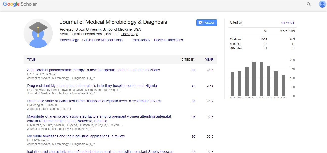
Medical Microbiology & Diagnosis received 14 citations as per Google Scholar report