Opinion - (2023) Volume 8, Issue 1
Received: 02-Jan-2023, Manuscript No. JPNM-23-90692;
Editor assigned: 04-Jan-2023, Pre QC No. P-90692;
Reviewed: 16-Jan-2023, QC No. Q-90692;
Revised: 21-Jan-2023, Manuscript No. R-90692;
Published:
28-Jan-2023
, DOI: 10.37421/2472-100X.2023.8.215
Citation: Rizaldi, Janicek. “An Overview of the Key Management Concerns for Paediatric Condylar Trauma.” J Pediatr Neurol Med 8 (2023): 215.
Copyright: © 2023 Rizaldi J. This is an open-access article distributed under the terms of the Creative Commons Attribution License, which permits unrestricted use, distribution, and reproduction in any medium, provided the original author and source are credited.
Children are not just "small adults," so treating them like adults can be inappropriate in many situations. As children grow and develop, their craniomaxillofacial (CMF) structure drastically shifts. The location, pattern, and nature of CMF injury are also altered by this anatomical change. In a similar vein, children's condylar architecture and anatomy are distinct, making the treatment of pediatric condylar fractures quite distinct from that of adult condylar fractures. A surgeon faces additional difficulties due to behavioral and physiological variations [1].
Condylar fracture in children may also benefit from conservative or nonoperative treatment. Paediatric facial growth, precise reduction, and rigid fixation are compromised by the choice between operative and non-operative management. Many factors influence this important decision. A child's facial growth and development can suffer greatly from improper treatment. It may result in ankylosis, among other deformities. As a result, pediatric condylar fracture treatment should be well-planned and carried out. Because children are not just "small adults," it can be inappropriate to treat them like adults in many situations. The structure of the craniomaxillofacial (CMF) area changes significantly as children develop. This anatomical change also changes the location, pattern, and nature of CMF injury. Similar to how children's condylar architecture and anatomy differ; pediatric condylar fracture treatment differs significantly from adult condylar fracture treatment. Behavioral and physiological differences present additional challenges for a surgeon.
Conservative or non-operative treatment may also be beneficial for children with condylar fracture. The choice between operative and non-operative management compromises paediatric facial growth, precise reduction, and rigid fixation. This crucial decision is influenced by numerous factors. Inappropriate treatment can have a significant impact on a child's facial growth and development. It could cause ankylosis, among other malformations. Therefore, treatment for pediatric condylar fractures ought to be meticulously planned and carried out [2].
Age also alters the condyle. The ramus and condyle's posterior borders show active growth while the anterior border shows resorption. The mandibular condyle's fracture patterns and distribution are influenced by the developmental anatomy of the lower jaw. In children between the ages of 2 and 5, the mandibular condyle is more likely to develop intracapsular comminuted fracture patterns. However, condylar neck fracture is the most frequently observed fracture pattern after the age of 5. This is due to the condyle's thin cortex and thickened periosteum at this age. Additionally, the condyle has a very thin neck. The paediatric condylar unit has high osteogenic potential due to its active growth centers and remodelling sites.
The adult mandibular condylar fracture is now typically treated with open reduction and internal fixation. However, ORIF is rarely used to treat pediatric condylar fractures. As a result, patients in this age range frequently receive non-operative treatment. The clinical outcomes of conservative (non-operative) management are typically satisfactory to excellent. Trauma to the condyle of a child or adolescent can cause growth disruption and long-term problems like occlusal problems, pain, masticatory problems, restricted mandibular movements, facial asymmetry, and debilitating conditions like temporomandibular joint disorders or ankylosis [3].
Two processes, endochondral replacement at the condyle and remodelling of the ramus, contribute to the increase in the vertical height of the ramus condyle unit. Both the mandible and maxilla develop at different rates of skeletal maturity. Girls reach skeleton maturity between the ages of 14 and 16, while boys do so between the ages of 16 and 18. However, growth may continue into the middle 20s. Because it matures later than the other facial bones, the mandible is more likely to sustain growth-related injuries. Therefore, the patient's age and the stage of mandibular growth have a significant impact on the fracture pattern.
Mandibular fracture is the most common facial bone fracture, followed by nasal bone fracture, accounting for 4–6% of all fractures1. The most common mandibular fracture is the condylar fracture. It accounts for between 30 and 40 percent of all mandibular fractures and between 11 and 16 percent of all facial fractures. Condylar fractures typically result from an indirect force impact on the chin or the body of the mandible rather than direct trauma. As a result, mandibular condylar fractures frequently go unnoticed. Imahara and colleagues found that mandibular fractures make up 32.7% of all facial fractures in children. Owusu and colleagues recently demonstrated that the condyle is the most frequently fractured site, with 14.6% cases. Of these, 20% have condylar fractures, 11.8% have condylar head fractures, and 9.4% have condylar base fractures. In any case, the component of injury and site of injury fluctuates with age and orientation [4].
When diagnosing condylar fractures in children, an accurate clinical and radiological evaluation is critical. The patient's age is a limiting factor in the diagnosis of a paediatric fracture. Eliciting subjective symptoms like inferior alveolar nerve dysfunction, pain, or malocclusion becomes difficult in younger patients. Condylar fractures are typically associated with symphysis, parasymphysis, and body fractures, so imaging remains the best method for supporting a diagnosis of a facial fracture in children, even as the patient becomes more responsive and cooperative with age. As a result, sublingual ecchymosis or laceration of the submental region may occur frequently. Due to the shortening of Ramal height on the ipsilateral side, the chin deflection to the affected side is a classic clinical feature. In such cases, open bite and flattening of the mandible's body can be seen on the opposite side [5].
Patients with bilateral condylar fractures may present with posterior gagging or occlusion and an anterior open bite. When ramal height is maintained despite the fracture, the occlusion, projection, and symmetry of the mandible can occasionally be maintained. When a young patient is likely to be uncooperative due to pain, taking a plain radiograph can be difficult because it requires patient cooperation. In addition, a straightforward radiograph may fail to reveal overlapping of the condyle-ramus complex. The most effective method for accurately diagnosing this region is computed tomography (CT), which may necessitate sedation in order to produce sufficient images free of movement artifacts.
None.
None.
Google Scholar, Crossref, Indexed at
Google Scholar, Crossref, Indexed at
Google Scholar, Crossref, Indexed at
Google Scholar, Crossref, Indexed at
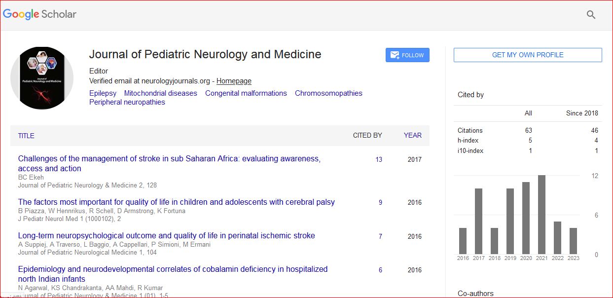
Journal of Pediatric Neurology and Medicine received 68 citations as per Google Scholar report