Review Article - (2022) Volume 12, Issue 8
Received: 04-Jul-2022, Manuscript No. MCCR-22-68382;
Editor assigned: 07-Jul-2022, Pre QC No. MCCR-22-68382(PQ);
Reviewed: 22-Jul-2022, QC No. MCCR-22-68382;
Revised: 03-Sep-2022, Manuscript No. MCCR-22-68382(R);
Published:
12-Sep-2022
, DOI: 10.37421/2161-0444.2022.12.189
Citation: Hussen, Narmin Hamaamin, Gashbeen
Osman Muhammed, Akar Yousif Yassin and Parwa Ahmed Esmail,
et al. "Anthracycline in Medicinal Chemistry: Chemistry,
SARs, Mechanism of Cardiotoxicity and Preventive Strategies." Med
Chem 12 (2022): 189.
Copyright: © 2022 Hussen NH, et al. This is an open-access article distributed under the terms of the creative commons attribution license which permits
unrestricted use, distribution and reproduction in any medium, provided the original author and source are credited.
Anthracyclines are among the most effective anticancer drugs ever developed. Cardiotoxicity is a well-known anthracycline side effect that restricts the total amount of medication given and can result in heart failure in some patients. Anthracyclines are thought to cause cardiotoxicity after one electron reduction with ROS overproduction or two electron reduction with conversion to C-13 alcohol metabolites, according to the pathophysiology of anthracycline induced cardiotoxicity. Overproduction of Reactive Oxygen Species (ROS) is likely to be the cause of anthracycline induced acute cardiotoxicity, but not all aspects of progressive cardiomyopathy. Secondary alcohol metabolites can play an important role in promoting cardiotoxicity's progression to end stage cardiomyopathy and congestive heart failure.
This review will provide an overview of the molecular mechanisms responsible for anthracycline induced cardiotoxicity focusing on the pathogenic role of Reactive Oxygen Species (ROS) and/or anthracycline secondary alcohol metabolites; and, lastly, on the most promising strategies to minimize or prevent anthracycline induced cardiotoxicity.
Anthracycline • Cardiotoxicity • ROS • Secondary alcohol metabolite • Prevention • Treatment
Cardiovascular diseases and cancer are the first and second leading causes of death in the world, respectively. When we consider the cardiovascular complications of anticancer therapies, these two conditions may become synergistic [1]. According to a report published by the national cancer institute in the United States, at least 16.9 million cancer survivors were alive in the United States in 2019, with that number expected to rise to more than 26 million by 2040 [2].
The development of efficient screening and treatment options for numerous neoplastic disorders during the last three decades has resulted in a large number of long-term cancer survivors. In five years, 67 percent of people diagnosed with cancer will be living, and in ten years, 75 percent of children diagnosed with cancer will be alive. In a seven years study of 1,807 cancer survivors, 33 percent died of cardiac disease and 51 percent died of cancer, according to the national health and nutrition examination [3]. Chemotherapy induced cardiotoxicity is caused mostly by anthracycline compounds, which are commonly used to treat lymphoma, sarcoma, breast cancer, and pediatric leukemia. Despite efforts to identify risk factors, create fewer toxic derivatives, and diagnose subclinical damage early, no consensus exists on the optimal strategy for preventing anthracycline induced cardiotoxicity.
Advances in the molecular basisof anthracycline induced cardiotoxicity may lead to more effective cardioprotective therapies in the future [4].
The purpose of this review is to provide an overview of research articles that have been published over the last 15 years (with few exceptions) on the chemistry, structure activity relationships, and molecular mechanisms underlying anthracycline induced cardiovascular toxicity, focusing on the role of pathogenesis in Reactive Oxygen Species (ROS) and/or secondary alcohol metabolites of anthracyclines; and finally, the most promising strategies for preventing or treating anthracycline-induced myocardial insufficiency (Figure 1).

Figure 1. Given figure discussing about chemistry and sars, anticancer, risk factors, treatments.
Anthracycline
Anthracyclines are a class of natural antibiotics that are among the most effective anticancer drugs now in clinical use, with only a few cancer forms resistant to them. Doxorubicin (DOX) and Daunorubicin (DNR) are natural anthracycline antibiotics that were originally isolated from Streptomyces peucetius var. caesius by Arcamone and Di Marco in the early 1960’s as part of an industrial company's methodical search for anticancer drugs (Farmitalia Research Laboratories of Milan). They were clinically tested shortly after and registered in the early 1970’s. They have been launched since then and have become the anthracycline class's prototypes. The first generation anthracyclines are still often prescribed and remain a mainstay in many chemotherapies schemes half a century after their development [5,6]. Doxorubicin is one of the most commonly used and effective chemotherapeutic agents. It is used as a first line treatment for a wide range of cancers, including lymphomas, sarcomas, and a wide range of solid tumors such as breast, lung, bladder, bone, and cervical malignancies in both children and adults [7,8]. While daunorubicin is used to treat acute lymphoblastic and myeloblastic leukemias [9].
DOX, DNR, Epirubicin (EPI), Idarubicin (IDA), pirarubicin, and amrubicin are the most common anthracyclines licensed for clinical use by the Food and Drug Administration (FDA) [10]. Combination therapy with anthracycline containing multidrug regimens that include doxorubicin, cyclophosphamide, and either docetaxel, paclitaxel, or fluorouracil achieves the best treatment outcomes [11].
Chemistry of anthracycline and structure activity relationship considerations
Anthracyclines share a tetracyclic aglycone structure consisting of four cyclohexane chains with a sugar moiety at carbon C-7 of ring A, adjacent quinone (hydroquinone) groups in rings B and C, a methoxy substituent carbon C-4 in ring D, a short side chain at (C-9) and a carbonyl group at (C-13) [12]. Doxorubicin is consisting of two different moieties. The aglyconic moiety consists of tetracyclic (anthraquinone) rings having a quinine hydroquinone adjacent group and a methoxy substituted short chain, as well as a short side chain with a carbonyl group at C-13 and primary alcohol at C-14. The second moiety is a daunosamine and consisting of a 3-amino-2,3,4-trideoxy-L-fucosyl moiety, which is connected to C-7 of ring A via a glycosidic bond [13-15]. A number of active sites including three functional groups: Ketone, amine and hydroxyl are present in the structure. Besides hydrophobic interactions possible through the rings, the functional groups allow electrostatic interactions such as hydrogen bonds to occur between DOX and other molecules [16]. Another extractive anthracycline, DNR, differs from DOX in that its side chain ends with a methyl group rather than primary alcohol. The side chain at C-9 differentiates doxorubicin from daunorubicin, the former holding an extra hydroxyl group at position 14. On an industrial scale, doxorubicin is currently made through semi-synthesis from daunorubicin by a chemical bromination at C-14, followed by bromine displacement by hydroxide under mild base treatment [17]. The hydroxylated tricyclic quinone system is the chromophore responsible for the red to the orange color characteristic of anthracyclines, which absorb around the 480 nm range of the visible light spectrum. Attached to ring A at the C-7 position is the unique glycone moiety 3-amino 2,3,6-trideoxy-α-Llyxo- hexopyranose, also known simply as daunosamine [18].
It is an unusual deoxy aminosugar conserved in tens of anthracycline congeners and considered crucial for anticancer activity because of the central role it plays in anthracycline’s principal mechanism of action.
Indeed, the daunorubicin aglycone, without the sugar moiety, shows decreased activity by 70-100 fold, compared to the parent compound.
Variation of the sugar core at the C-3ʹ amino position, including disubstituted compounds at C-3ʹ , with an equatorial methyl group and axial hydroxy or methoxy groups, led to complete loss of activity, while the presence of a monosubstituted equatorial methoxy group at the same C-3ʹ position gave rise to a derivative with 3-fold lower activity than daunorubicin, as judged by cytotoxicity on colon cancer SW620 cells. These modifications with different substituent patterns showed that 2,6-dideoxy sugars should be preserved for optimal potency [19,20]. The essentiality of daunosamine is no surprise since this typical structural unity can be found in many other bioactive glycosidic natural products, including antibiotics from the macrolide, polyene, and glycopeptide classes, such as erythromycin A, amphotericin B, and vancomycin, respectively (Figure 2).

Figure 2. Structure of the anthracycline drugs and structureactivity relationship.
Mechanism of anticancer activity of anthracycline
The cytotoxic activity of anthracyclines is due to a strong inhibitory effect on nucleic acid synthesis, but the specific molecular mechanisms of action are still being debated. It is now well accepted that anthracyclines work through a variety of mechanisms, the most consistent of which are, intercalation into DNA, oxidative stress induction, and, most crucially, poisoning of the Topoisomerase II enzyme (Top II). The ability of anthracyclines to connect with DNA as a result of intercalation of their planar ring system strengthened by auxiliary binding of the amino sugar was the main mechanism by which they demonstrate their cytotoxic effects. In the minor groove, the sugar moiety interacts with ring A and inhibits topoisomerase II by stabilization of the cleavable complex. The amino sugar seems to play a very important role and compounds lacking this functionality failed to inhibit the enzyme. But for the doxorubicin and daunorubicin, specificity is provided by the hydrogen bonding between O-9 of the anthracycline and N-2 and N-3 of guanine. The formation of covalent bonds between anthracyclines and DNA also is supported by several studies in which formaldehyde is produced by oxidation of cellular components or other anthracycline molecules. This oxidation results from the production of ROS such as H2O2, which are generated during the redox cycling of anthracyclines. The generated formaldehyde may then form a methylene bridge between the 4-amino group of the anthracycline and the 2-amino group of guanines in DNA (Figure 2). Modifications at C-3ʹ , such as acylation of the amino group may reduce cytotoxicity, replacing the amino function with a small group, such as hydroxyl, can prevent multidrug resistance while maintaining the cytotoxic effect. The method of binding to DNA and Top II is altered by other structural modifications, such as the inversion of configuration from the anomeric location of daunosamine.
According to conformational and crystallographic studies, daunosamine is the most flexible domain in anthracyclines, capable of adopting a variety of stable conformations for an optimal fit of the sugar at the DNA topoisomerase II interface. As a result, amino sugar changes are not only tolerated in terms of bioactivity, but also of potential interest in medication development. The anthracyclines are considered to be specific for the S phase of the cell cycle. There are many data to support increased binding of p53 with DNA when anthracyclines are administered, stimulating the apoptotic process, whereas some studies have failed to confirm this Figure 3.

Figure 3. Formation of anthracycline DNA adduct.
Risk factors for cardiotoxicity induced by anthracyclines
The cytotoxic effects produced by anthracyclines are not limited to tumor cells. Anthracycline based chemotherapy can have significant and dose limiting side effects on normal tissue. Anthracyclines' therapeutic potential is restricted by their selective toxicity for cardiac tissue, which can result in progressive, chronic cardiomyopathy that progresses irreversibly and unpredictably to Congestive Heart Failure (CHF). This is most evident in the case of daunorubicin and doxorubicin and less problematic with the newer derivatives idarubicin and epirubicin. Cardiovascular problems were first documented a few years after daunorubicin was introduced. In 1979, a dose toxicity curve was constructed by plotting the incidence of heart failure (defined by clinical signs and symptoms such as shortness of breath, jugular vein dilatation, S3 gallop, cardiomegaly, hepatomegaly, or pericardial effusion) versus total anthracycline dose used in many studies. In a retrospective study, Von Hoff et al.
discovered that when a patient receives a cumulative dose of doxorubicin of 400, 550, or 700 mg/m2, the incidence of cardiotoxicity is 3, 7, and 18 percent, with dose limiting toxicity, respectively. As a result, it is recommended that the total doxorubicin dose not exceed 550 mg/m2. If epirubicin is used, the maximum cumulative dose should not exceed 900 mg/m2. Ryberg et al. conducted a risk analysis of 1,097 patients with metastatic breast cancer who had previously received epirubicin treatment; the maximum cumulative dose for epirubicin treatment was determined based on the patient's risk level, with the goal of keeping the incidence of congestive heart failure below 5%. According to the findings, the recommended cumulative dose for patients under the age of 40 with no other risk factors for congestive heart failure is 806 mg/m2, while the maximum cumulative dose for people over the age of 70 is 609 mg/m2. A cardiac biopsy was performed to assess cardiac damage in anthracycline treated individuals, with morphological changes graded using the Billingham system. The degree of myofibrillar loss or vacuolization is used to determine the severity of cardiotoxicity in this system. With a cumulative dose of as little as 200 mg/m2, some patients showed morphologic abnormalities. However, due to its invasiveness, a cardiac biopsy is not regularly performed in today's practice. Detecting troponin leakage in the peripheral blood during or after anthracycline treatment was found to be a good, less intrusive alternative to biopsy for detecting cardiac harm caused by anthracycline treatment. There is no safe cut-off point for anthracycline induced cardiotoxicity because troponin can be detected in some patients as early as the end of the first anthracycline delivery cycle. Early or late-onset anthracycline induced cardiomyopathy has been characterized using a one year threshold following anthracycline treatment. After one year of anthracycline treatment, the cumulative incidence of cardiac incidents peaked.
Many other risk factors may increase the incidence and severity of anthracycline related cardiotoxicity, depending on patient and treatment features. Age at treatment (less than 18 or more than 65 years), sex (female), race (black), genetic polymorphisms, preexisting cardiovascular pathologies (coronary artery disease, left ventricular dysfunction, hypertension, etc.), metabolic (diabetes), or genetic (trisomy 21) diseases, as well as higher administration rates, concomitant radiation therapy, and combination chemotherapy have all been shown to increase the risk of developing anthracycline induced cardiotoxicity.
Molecular mechanisms of anthracycline-induced cardiotoxicity
The mechanism of anthracycline induced cardiomyopathy is still debated and understudied. Various molecular pathways have been postulated to explain anthracycline cardiotoxicity throughout the years, but no one explanation appears to include all of the known fundamental and clinical data. The current theory is that anthracyclines are harmful in and of themselves, but that following one electron reduction through ROS overproduction or two electron reduction through conversion to C-13 alcohol metabolites, they become even more lethal. ROS appears to be responsible for early cardiac abnormalities, whereas secondary alcohol metabolites may play a role in boosting cardiotoxicity's progression to end stage cardiomyopathy and CHF. Furthermore, "acute" toxins such as ROS may lead to increased levels of "chronic" toxins such as secondary alcohol metabolites, eventually priming the heart to develop cardiomyopathy and CHF, according to a unifying hypothesis.
Role of reactive oxygen species in anthracycline induced cardiotoxicity
Anthracyclines can generate ROS generation in two ways: through an enzymatic mechanism involving several mono-electronic oxidoreductases and through a non-enzymatic mechanism involving anthracycline complexes iron. Several enzymes, including NAD (P)Hoxidoreductases and CYP reductases, reduce the C ring of anthracyclines by one or two electrons, resulting in semiquinone or hydroquinone. In the enzymatic mechanism, one-electron reduction of the quinone moiety in the C ring of anthracyclines results in the formation of a semiquinone radical, which quickly regenerates its parent quinone by reducing molecular oxygen to superoxide anion (O2•) and its dismutation product Hydrogen Peroxide (H2O2), thus increasing ROS concentration above physiologic levels generated during normal aerobic metabolism. The radical is normally converted to H2O2 by superoxide dismutase and H2O2 is then converted to H2O and O2 by catalase. Alternatively, ROS production may also occur via a non-enzymatic pathway triggered by anthracycline-iron complexes.
However, the production of superoxide (O2-•) is also associated with the release of iron from intracellular stores, which may be chelated by anthracycline. Anthracyclines indeed bind avidly to Fe3+, forming a drug-metal complex which, in the presence of oxygen, can cycle between the Fe2+ and Fe3+ oxidation states. The iron then causes catalase to divert the normal detoxification pathway, resulting in the production of more active radicals such as the Hydroxyl radical (OH).
Catalase levels in myocardial cells are lower, making them less able to detoxify the H2O2 produced by redox cycling. As a result of the elevated levels of OH, for which there is no detoxification pathway, cellular damage occurs. Reactive oxygen species, or ROS, are materials like O2-, OH, and H2O2 cause cellular damage when present in high enough concentrations. Single-strand breaks in DNA are caused by hydroxyl radicals, which activate p53 and increase apoptosis in cardiac cells. Doxorubicin administration has also been shown to activate both the intrinsic and extrinsic pathways of apoptosis in a variety of ways, which has been linked to an increase in H2O2 and hydroxyl radical production. One proposed mechanism involves damage to the mitochondrial membrane by H2O2 and OH resulting in the release of cytochrome C, which would activate the intrinsic pathway. Anthracycline aglycones may generate ROS as well. Due to their higher lipophilicity, aglycones can intercalate into mitochondrial membranes better than the parent anthracyclines and thus divert more electrons towards oxygen thereby generating ROS in the closest proximity to sensitive targets. In most cells, ROS and RNS formation is kept to a minimum by the presence of detoxifying enzymes, like superoxide dismutase, catalase, and glutathione peroxidase. Unfortunately, cardiomyocytes are more susceptible than other cells to oxidative damages for several reasons, namely:
• High metabolic activity.
• High concentration of cardiolipin, a mitochondrial membrane bound phospholipid critical to cell respiration for which anthracyclines have a high affinity.
• Weak antioxidative defenses due to their low content of catalase, superoxide dismutase, and glutathione peroxidase. Furthermore, DOX administration actually diminishes superoxide dismutase and glutathione peroxidase activity.
However, the role of oxidative stress in anthracycline-induced cardiotoxicity is becoming increasingly under question because most of the evidence favoring this hypothesis has been obtained from subcellular fractions with extremely high drug concentrations never employed in clinical practice (Figure 4). Although ROS overproduction should probably be taken into account for several aspects of anthracycline acute cardiotoxicity, it cannot fully explain progressive cardiomyopathy. In fact, it has been demonstrated that:

Figure 4. Process of anthracycline redox cycling.
• Anthracyclines at therapeutic concentrations do not succeed in producing ROS.
• Anthracycline cardiotoxicity currently develops in the absence of biochemical markers of oxidative damage, and, unexpectedly, DOX decreases the myocardial release of conjugated dienes and hydroperoxides.
• ROS scavengers and/or antioxidants, that afford protection in small animals, fail in preventing or decreasing chronic cardiac dysfunction in large animals or in patients treated with multiple doses of anthracycline.
Role of secondary alcohol metabolites in anthracycline induced cardiotoxicity
Additional cardiotoxicity mechanisms have been suggested, including the metabolic reduction of the anthracycline C-13 ketone to alcohol. Several pharmacokinetic, functional, and clinical studies suggest the role of secondary alcohol metabolites in the pathophysiology of anthracycline induced cardiomyopathy. After so many DOX injections in animal studies, DOXol but not DOX accumulates exclusively in the heart, which appears to represent intramyocardial anthracycline metabolism rather than metabolite uptake from the flow. In DOX treated patients, human post-mortem investigations show that the heart is one of the organs with the highest concentration of DOXol, the most abundant anthracycline metabolite maintained inside cardiomyocytes. These findings imply that DOXol has a lower clearance in cardiomyocytes than DOX or a polar aglycone and develops a long-lasting reservoir, which could explain the time delayed character of anthracycline cardiotoxicity and the heart's unique susceptibility to this family of chemotherapeutic drugs. The resulting alcohols have been linked to a variety of pharmacological effects, including loss of Ca+2 homeostasis and inhibition of Na+/K+-ATPase in cardiac cells. The formation of doxorubicin C-13 alcohol, doxorubicinol has been associated with the conversion of Iron Regulatory Protein-1 (IRP-1) into a null protein that is no longer able to maintain iron homeostasis. It's been suggested that doxorubicin induced cardiotoxicity is caused by a lack of iron regulation. From a mechanistic viewpoint, secondary alcohol metabolites are several fold (~20 to 40 times) more potent than their parent anthracyclines at inhibiting the Ca2+/Mg2+- ATPase of the sarcoplasmic reticulum, DOXol is also more effective than C-13 deoxy DOX (a DOX analog missing the C-13 carbonyl moiety and therefore unable to form DOXol) at suppressing the gene expression of the sarcoplasmic reticulum's Ca2+ transporting ATPase (SERCA2) and cardiac calcium release channel (RYR2). Daunorubicin and idarubicin, on the other hand, form more alcohol metabolites than doxorubicin and have similar and reduced cardiotoxicity, respectively.
This indicates that cardiotoxicity is caused by more than just alcohol metabolism (Figure 4). Despite their high cardiotoxicity, anthracycline 13-hydroxy metabolites have a slight (or no) antitumoral impact.
DOXol and DNRol, are slightly less effective than the parent anthracyclines at inhibiting topoisomerase and destroying tumor cells.
As a consequence, cytosolic reductases appear to be a significant factor in the pathophysiology of anthracycline-induced cardiomyopathy and the development of tumor resistance to anthracyclines. Studies with transgenic mice that overexpress human carbonyl reductase solely in the heart indicate that DOXol levels in expressers are four times greater than in nonexpressers.
Furthermore, expresser mice develop cardiomyopathy more quickly than nonexpresser mice and die much sooner following DOX treatment. In contrast, mice carrying a null allele of the Carbonyl Reductase 1 (CBR1) gene (Cbr1+/) have lower plasmatic levels of DOXol and a lower incidence of anthracycline-induced cardiomyopathy than mice carrying two active genotypes (Cbr1+/+).
The human CBR1 gene is located on chromosome 21, and people with down syndrome have three copies of it. High quantities of human CBR1 mRNA are found in down syndrome cells, which may help to explain the enhanced chemotherapy toxicity seen in down syndrome patients. Studies looking into the molecular basis of the cardiotoxic synergism between DOX and Paclitaxel (PTX), the first approved member of a family of nonanthracycline chemotherapeutics known as taxanes, have also confirmed the importance of secondary alcohol metabolites in the development of anthracycline induced cardiomyopathy. Both DOX and PTX are quite active in breast cancer. Finally, novel anthracycline analogs lacking the side chain carbonyl group that is susceptible to reduction to toxic secondary alcohol metabolites (e.g., C-13 deoxyDOX) or have reduced affinity for cardiac cytosolic reductases (e.g., EPI) exhibit a less severe effect in rodents. When C-13 deoxyDOX was tested in humans, cardiotoxicity was noted, although the cumulative dose was quite high. Apart from casting doubt on the involvement of anthracycline alcohol metabolites, these findings call for more research into the pharmacokinetics and cardiotoxicity of this DOX group (Figure 5).

Figure 5. Schematic route metabolism of doxorubicin.
Anthracycline induced cardiotoxicity: A unifying mechanism
ROS overproduction and the formation of C-13 alcohol metabolites may be related and synergistic in the development of anthracyclineinduced cardiomyopathy (Figure 3). The quinone group is reduced by one electron, creating the semiquinone radical. The starting quinone is rapidly regenerated by reducing molecular oxygen to the superoxide anion (O2), and the latter is dismutated to produce Hydrogen Peroxide (H2O2). As a result, an extended redox cycle of the quinone moiety exposes cardiomyocytes to Reactive Oxygen Species (ROS), which when combined with iron can produce oxidative stress. The upregulation of oxidative stress can be particularly damaging to the heart when cardiomyocytes lack oxyradical scavenging enzymes such as superoxide dismutase and catalase or glutathione peroxidase production of cardiac aldo-keto reductases by the generation of H2O2 and by-products of lipid peroxidation (such as malondialdehyde and 4-hydroxynonenal).
Increased production of dangerous anthracycline metabolites is associated with increased synthesis of these enzymes, allowing cardiomyocytes to detoxify lipid aldehydes while enhancing anthracycline conversion to secondary alcohol metabolites, leading to heart damage. Finally, oxidative stress caused by anthracycline oneelectron reduction and semiquinone redox may be a precursor to chronic progressive cardiomyopathy caused by anthracycline C-13 alcohol metabolites. In addition, when the carbonyl group is reduced by two electrons, a secondary alcohol metabolite is formed that is much more polar than the parent anthracycline. As a result, while anthracyclines are easily removed from cardiomyocytes, anthracycline secondary alcohol metabolites have little cardiac clearance and tend to build a toxic anthracycline reservoir. Long-lived anthracycline metabolites contribute to oxidative damage but also produce toxicity through mechanisms other than oxidative stress, such as the inhibition of many ATPases or the activation of iron regulatory proteins that control cellular iron absorption and storage (Figure 6).

Figure 6. Unified mechanism for anthracycline induced cardiotoxicity.
Semiquinone is produced when the quinone molecule undergoes one-electron redox cycling, turning oxygen into ROS. The development of cardiotoxicity has been attributed to further ROS interactions with iron. In contrast to parent anthracyclines, a secondary alcohol metabolite is produced when the side chain carbonyl group undergoes a two electron reduction. This substance accumulates to build a long-lasting anthracycline reservoir that is also associated with cardiotoxicity. Modified from: ROS, Reactive Oxygen Species; DOX, Doxorubicin; DOXOL, Doxorubicinol (secondary alcohol metabolite).
Strategies for preventing and treating anthracycline induced cardiotoxicity
To prevent or treat cardiotoxicity associated with anthracyclines, multiple approaches have been developed including:
Prevention of cardiotoxicity: To maximize the beneficial effects of anthracyclines, much effort has been spent devising new strategies to decrease or prevent their cardiotoxic effects.
Limiting cumulative dose: Maximum doses of anthracyclines have been introduced. Maximum cumulative doses of 400-550 mg/m2 doxorubicin and 900 mg/m2 epirubicin are currently recommended.
Prolonging administration time: Weekly low doses and a long continuous infusion time (24-96 h) help to reduce cardiac toxicity. A study indicated that giving doxorubicin intravenously over a long period of time (>6 hours) reduced the incidence of clinical heart failure and subclinical myocardial injury. The weekly medication had lower cardiotoxicity than the normal three-week treatment.
Strengthening the monitoring of cardiotoxicity: The monitoring of cardiotoxicity during the use of anthracycline medicines should be enhanced to achieve early diagnosis and treatment, preventing severe cardiotoxicity. There are several approaches for monitoring cardiac toxicity that have been reported: Cardiac endocardium biopsy, electrocardiogram, echocardiography, myocardial backscatter integration parameters, tissue doppler imaging, ischemic modified albumin, troponin, and brain natriuretic peptide. These monitoring methods have certain limitations, such as invasiveness and low specificity. Therefore, continuous efforts are needed to find sensitive, specific, and non-invasive monitoring techniques and tools.
Development of liposome anthracyclines: High peak plasma levels of anthracyclines may contribute to cardiotoxicity. In addition to various dosing schedules aimed at lowering peak plasma concentrations, an alternative strategy has been liposomal encapsulation. Liposomes are fat spheres containing details.
Liposome anthracycline encapsulates the drug in a lipid body to protect it from degradation and inactivation in the plasma, and it takes advantage of structural defects in the tumor tissue, such as vascular endothelial discontinuity and lymphatic vessel breakage, to allow anthracycline drugs to selectively penetrate the vascular system and enter the tumor tissue. This lowers the drug's concentration in normal endothelium tissue, which can significantly decrease the toxicity to the heart while enhancing the anti-tumor index and expanding the anti-tumor spectrum (Figure 7).

Figure 7. Active targeting using doxorubicin encapsulated liposomes with functionalized surfaces.
Two main agents include a pegylated formulation called Doxil® (Ben Venue Laboratories, Johnson and Johnson) and a second agent called TLC D-99 or Myocet® (Elan Corporation, Plc). Phase II trials suggest that liposome encapsulated doxorubicin combination therapy is highly active against metastatic breast cancer and is less cardiotoxic than doxorubicin therapy alone. The most commonly used formulation is pegylated liposomal doxorubicin (Doxil) was approved in 1995. The pharmacokinetics of medications contained within pegylated liposomes are altered. These drugs have a long circulation half-life as they resist uptake by macrophages and monocytes.
Furthermore, liposomes have the ability to extravasate through leaky tumor vasculature and directly localize large amounts of doxorubicin at tumor sites. This localization was first observed in highly vascular tumors, such as kaposi’s sarcoma, in which liposomal doxorubicin was more effective than the combination of standard doxorubicin, bleomycin, and vincristine. Pegylated liposomal doxorubicin has been evaluated in several phase III trials as a first line treatment of metastatic breast cancer. Pegylated doxorubicin has also been studied in the treatment of recurrent epithelial ovarian cancer, with efficacy similar to conventional therapy and a better safety profile.
Pegylated liposomal doxorubicin was less toxic to the heart but just as effective in terms of progression free and overall survival.
Pegylated doxorubicin has a lower tendency to accumulate in myocardial cells, which reduces cardiotoxicity. Pegylated doxorubicin has lower free drug concentrations in the plasma and a limited concentration in cardiac cells than standard doxorubicin. This improved cardio-sparing impact has been reported in two studies that used endomyocardial biopsies to detect anthracycline induced cardiac damage. Cardiac changes in patients given pegylated doxorubicin are less severe than those in patients given similar cumulative doses of standard doxorubicin. Serial measurements of LVEF and clinical follow-up indicate that the risk of cardiomyopathy is significantly less than that of standard doxorubicin at cumulative doses of >500 mg/m2. Recent studies have shown that pegylated doxorubicin in conjunction with trastuzumab is not associated with the high levels of cardiotoxicity or HF seen in traditional doxorubicin and trastuzumab combination therapy. At least three randomized controlled trials with promising cardioprotective results have been conducted with the non-pegylated liposomal anthracycline formulation (Myocet). In addition to doxil and myocet, there are various liposomal anthracycline formulations. A non-pegylated form of liposomal doxorubicin has been approved in europe for use in metastatic breast cancer, but it is still under investigation in the US.
The use of these agents has been limited because of their high cost and negligible extracardiac toxicity.
Cardioprotective agents: Another strategy for preventing anthracycline induced cardiotoxicity is to combine cardioprotective agents with anthracyclines to reduce their cardiotoxic effects. This approach implies that cardiac damage and anti-tumor activity have separate mechanisms. With variable degrees of success, a number of distinct classes of agents have been investigated for their cardioprotective effects in the presence of anthracyclines.
Dexrazoxane (also known as ICRF-187) is currently the only FDA approved medication for the prevention of anthracycline-induced cardiotoxicity (licensed in the united states and canada as zinecard® and in europe as cardioxane®). Dexrazoxane has been proven to be effective in reducing both acute and chronic cardiotoxicity induced by anthracycline therapy. Dexrazoxane is a bisdioxopiperazine compound that is the hydrolysis of its piperazine rings, releases a diacid diamide (code-named ADR 925) that is an analog of e iron chelator Ethylene Diamine Tetra-Acetic Acid (EDTA). Unlike EDTA, dexrazoxane easily passes into cells. It was developed as an antitumor agent and is the (+) -(S)-enantiomer of racemic razoxane. Dexrazoxane is an intravenous bidentate chelating medication that binds intracellular iron and inhibits the generation of iron dependent free radicals. Because of their weakened intracellular defenses, myocytes are particularly vulnerable to oxidative damage.
Dexrazoxane binds to intracellular iron and prevents the conversion of superoxide anions and hydrogen peroxide to super hydroxide free radicals. It also prevents ferrous iron from being converted to ferric iron for utilization by superoxide anions. As a result, the free iron pool is decreased in the myocardium, and iron is prevented from binding with anthracyclines and damaging cells. Dexrazoxane can also change the configuration of topoisomerase 2, preventing anthracyclines from binding to it and thereby reducing cardiomyocyte death, mitochondrial dysfunction, and anti-oxidant gene suppression.
Dexrazoxane is a topoisomerase II inhibitor that, unlike anthracyclines, does not cause strand breaks. The clinical dose for cardiac protection is 10:1 to 20:1 of the doxorubicin dose. The American Society of Clinical Oncology (ASCO) guidelines for dexrazoxane advocate its use during the treatment of metastatic breast cancer when the cumulative doxorubicin dose exceeds 300 mg/m2 or in individuals with metastatic breast cancer who have received doxorubicin 300 mg/m2 and who will continue to receive doxorubicin therapy to maintain tumor control. In the US, dexrazoxane is approved for cardiac protection in women with metastatic breast cancer who have received 300 mg/m2 of anthracyclines or more. In Canada, it is approved for patients with metastatic breast cancer after 150 mg/m2 of anthracycline, and in europe, several countries have approved it for both anthracycline cardioprotection and for treating anthracycline extravasation. In one study, children with leukemia were treated with a cumulative dose of doxorubicin 300 mg/m2 and were randomly assigned to receive either doxorubicin alone or a dose of dexrazoxane before each doxorubicin dose. Serum cardiac troponin-T levels were measured as surrogate markers of cardiac injury. Children treated with doxorubicin alone (n=101) were more likely to have elevated troponin-T levels than were those who received dexrazoxane and doxorubicin (n=105; 50 versus 21%; p<0.001), and the groups had similar survival rates. It is unknown whether dexrazoxane can prevent chronic progressive cardiomyopathy after anthracycline treatment. Long-term follow-up research will be critical in answering this question. Treatment of pediatric Hodgkin’s disease with dexrazoxane added to doxorubicin, bleomycin, vincristine, and etoposide (ABVE) and dose intensified ABVE with prednisone and cyclophosphamide (ABVE-PC), followed by low-dose radiation, may be associated with an increased incidence of second malignant neoplasms (4 years standardized incidence rate: 41.86 versus 10.08%; p=0.02) and of acute myeloid leukemias/myelodysplastic syndromes (4 years standardized incidence rate: 613.6 versus 202.4%; p=0.10). This is in contrast to childhood acute lymphoblastic leukemia, for which similar lengths of follow-up found that rates of second malignancies were not increased by dexrazoxane, suggesting that if there is an effect in childhood Hodgkin's disease survivors, it may be related to disease or protocolrelated factors. Moreover, dexrazoxane can be responsible for different adverse effects such as a reversible elevation of hepatic transaminases as well as some myelotoxicity (neutropenia and thrombocytopenia), limiting the dose given to the patient.
New generation anthracycline drugs: Researchers have been looking for a "better anthracycline" with the same (or even greater) anticancer effectiveness but minimal or no cardiotoxicity for almost 30 years. Thousands of analogs have been identified or synthesized in the search for superior anthracyclines that could overcome the cardiotoxicity and resistance limitations of the original drugs. The tetracyclic rings have changed over the last decades, Figure 8 shows the side chain and the amino sugar. The original rationale was to prevent significant changes in the general molecular structure and chemical properties and to maintain the structural requirements for the activity that led to epirubicin and other second generation anthracyclines such as idarubicin, which have shown clinical efficacy and are the best known derivatives.

Figure 8. Dexrazoxane has a dual mode of action that allows it to chelate iron through the diacid diamide product of hydrolysis or avoid cardiotoxicity by blocking topoisomerase II, which is constitutively produced in cardiomyocytes.
Second-generation anthracyclines: Epirubicin, (compound 1, Figure 9) a doxorubicin epimer, is less likely to cause cardiotoxicity than doxorubicin. It has similar efficacy as doxorubicin, but it has fewer toxic side effects, resulting in a higher therapeutic index and therapeutic ratio, allowing for higher dosages and a wider margin of safety. In fact, the recommended maximum cumulative dose of epirubicin is 900-1000 mg/m2. The 900 mg/m2 cumulative dose of epirubicin is oncologically equivalent to doxorubicin ˜600 mg/m2, and it has the cardiotoxicity risk of doxorubicin ˜450 mg/m2. One randomized study found that the median cumulative dose at which HF occurred was epirubicin ˜1100 mg/m2 and doxorubicin 500 mg/ m2. However, a large retrospective study of patients with metastatic breast cancer treated with epirubicin found a 15%incidence of HF at doses of >950 mg/m2 and noted an additive effect of radiation to the risk of developing marked cardiotoxicity. Epirubicin has been linked to subclinical heart damage in several other investigations, even at low doses. A randomized, double blind study comparing epirubicin to doxorubicin demonstrated no clinically significant cardiotoxicity during epirubicin treatment, and no patients receiving epirubicin had HF at cumulative doses of 400-500 mg/m2. However, at a mean cumulative dose of 450 mg/m2, LVEF deteriorated considerably in a third of individuals. A subsequent study confirmed that relatively low doses of epirubicin result in myocardial injury, as indicated by decreases in LVEF and increases in natriuretic peptide levels. Whether these abnormalities increase the long-term risk of HF years after therapy is unknown.

Figure 9. Second and third generation anthracyclines. 1: Epirubicin, 2: Idarubicin, 3: Valrubicin, 4: Amrubicin, 5: Pirarubicin, 6: Aclarubicin, 7: Sabarubicin, 8: Nemorubicin
Idarubicin, (compound 2, Figure 8) a synthetic derivative of daunorubicin, is more lipophilic and has a longer half-life than daunorubicin. Idarubicin is effective at treating lymphoid and myeloid leukemias, non-Hodgkin’s lymphoma, and advanced breast cancer.
Similar to epirubicin, idarubicin causes less cardiotoxicity than doxorubicin. A study of patients with leukemia treated with idarubicin found that the incidence of cardiomyopathy was 5% at a cumulative dose of 150-290 mg/m2. Again, sub-clinical cardiac effects were observed even at lower cumulative doses: at a cumulative dose of just 150 mg/m2, the incidence of a marked decrease in LVEF (a change in LVEF ≥ 10%, resulting in a final level of ≥ 50%) was 18%, and the incidence of a moderate or greater asymptomatic decrease in LVEF (a change in LVEF ≥ 15% to a final level ≤ 45%) was 7%. Therefore, although HF is uncommon with cumulative doses of idarubicin <300 mg/m2, asymptomatic echocardiographic changes are more frequent.
The predictive value of these echocardiographic abnormalities for the development of future HF is unclear.
Valrubicin, (compound 3, Figure 9) a semisynthetic counterpart of doxorubicin is prepared from 14-iodo-N-trifluoroacetyldaunorubicin and sodium valerate or by enzymatic acylation of 14-iodo-Ntrifluoroacetyldaunorubicin with valeric acid. Valrubicin increases cell penetration and concentration in the perinuclear cytoplasm because it has higher lipophilicity than doxorubicin. This modulation of cytoplasmic protein kinase C activity culminates in the induction of death. Like other anthracycline venoms, the metabolite targets nuclear topoisomerases after being converted to ntrifluoroacetyldoxorubicin. However, its activity is somewhat inhibited by conventional resistance mechanisms carried out by efflux transporters, to which it undergoes deacylation. It was clinically designed to be delivered via intravesical instillation to treat bladder cancer, despite being less cardiotoxic than doxorubicin with systemic administration (FDA registered). Only medication licensed for individuals with bladder cancer in situ who are bacillus calmette Guerin refractory capable of having a radical cystectomy.
Amrubicin, (compound 4, Figure 9) is a fully synthetic drug, analogue of anthracyclines created through simplification. It is a simplified form of daunosamine that differs from daunorubicin in that it does not contain the 4-methoxy group of the aglycone or the 3- amino group of the carbohydrate. In contrast, at position 9 of the structure, an amino group takes the place of the hydroxyl group. It is a prodrug made active by enzymatic conversion of the side chain carbonyl group to a hydroxyl (13-OH) group. In contrast to the secondary alcohol metabolites of other anthracyclines, the active metabolite stabilizes the cleavable complex of topoisomerase II with DNA and inhibits cell growth up to 200 times more than the parent substance. Amrubicin's primary toxic effect is myelosuppression, making it less cardiotoxic than doxorubicin and epirubicin.
Although it was licensed for use in japan in 2002 to treat small-cell and non-small cell lung cancers, clinical trials are still being conducted to test its efficacy as a first and second line therapy for lung cancer and other cancers. Few other analogues made it into clinical development despite extensive testing, and even fewer got the green light for sale.
Semi-synthetic pirarubicin (compound 5, Figure 9), also known as 4-tetrahydropyranyldoxorubicin, is active against some doxorubicin resistant cell lines and is thought to be less cardiotoxic than doxorubicin.
Aclarubicin (compound 6, Figure 9), compared to doxorubicin and daunorubicin, is said to have less cardiotoxicity, although it only slightly improves drug resistance. This naturally occurring anthracycline was obtained from Streptomyces galilaeus and has a distinctive oligosaccharide glycone moiety and a variant aglycone structure with a carboxyl ester at C-10. Aclarubicin works through a variety of methods, including reducing mitochondrial respiratory activity, blocking Top-II prior to DNA damage, and particularly histone eviction from nucleosomes, which results in alterations to the transcriptome. These medications are still not widely used worldwide and have just recently been registered in a few nations.
Third-generation anthracyclines: Regarding the third-generation anthracyclines being tested in clinical trials, some of them are showing promise as therapeutic candidates.
Sabarubicin, MEN 10755 (compound 7, Figure 9) derives from the glycosylation of 4-demethoxydoxorubicinone with a disaccharide, resulting in a glycoside in which a 2,6-dideoxy--L-lyxohexopyranosyl residue is positioned between the aglycone and daunosamine is. This differs from the natural anthracycline disaccharides, which have the amino sugar as the first moiety attached directly to the aglycone. The extended glycone allows the second sugar to interact both at the minor groove and with a DNA base. It is less cardiotoxic than doxorubicin but does not show the ability to overcome transport mediated resistance. In comparison to DOX, sabarubicin has a wider range of antitumoral activity as well as lower cardiotoxicity.
Furthermore, unlike DOX, sabarubicin histologic and functional cardiotoxic symptoms are not progressive. The decreased cardiac uptake of sabarubicin compared to DOX or EPI is now due to a rare combination of pharmacokinetic (reduced cardiac uptake), biochemical (reduced conversion to alcohol metabolite), and pharmacodynamic (reduced reactivity of the alcohol metabolite with aconitase/IRP-1) determinants. The cardiotoxic potential of MEN 10755 is also reduced by its impaired conversion to the secondary alcohol metabolite MEN 10755ol, according to studies with human cardiac cytosol and isolated rat heart (MEN 10755 forms 50 percent less alcohol metabolite than DOX.
Nemorubicin (3-deamino-3-[2-(S)-methoxy-4-morpholinyl] doxorubicin; PNU152243A; MMDX) (compound 8, Figure 8) is an investigational anthracycline in phase II/III clinical trials for hepatocellular carcinoma intrahepatic artery chemotherapy. MMDX is a DOX derivative with a methoxy morpholinyl group in the sugar moiety at position 3'. It has been chosen for the clinical study due to its efficacy against multidrug resistant cancers in vitro and in vivo, as well as the lack of cardiotoxicity at therapeutic doses. In vivo, MMDX is 80-150 times more potent than DOX, but in vitro, the drug is only 2-10 times more potent than DOX against tumor and hematopoietic cells. MMDX's cardiac safety and therapeutic effectiveness would undoubtedly need further investigation.
Treatment of cardiotoxicity
In the early detection of anthracycline induced cardiotoxicity symptoms, such as mild arrhythmia, atrial fibrillation, pericarditis, etc., anthracyclines should be discontinued by switching to a chemotherapy regimen without anthracyclines, and the necessary symptomatic treatment should be given. When patients have symptoms of congestive heart failure, anti-heart failure treatments, such as Angiotensin Converting Enzyme Inhibitors (ACEI), β blockers, calcium channel blockers, statins, aldosterone antagonists and combination therapy can be given.
Angiotensin Converting Enzyme Inhibitors (ACEI): ACE inhibitors, such as captopril and enalapril, have been used extensively in the non-cancer setting to reduce afterload in patients with ventricular dysfunction. They are now first-line medications in managing patients with symptomatic and asymptomatic LV dysfunction. In an early study, it was concluded that zofenopril has a complete protective effect on cardiomyocytes from chronic cardiomyopathy caused by doxorubicin, without affecting the antitumor activity of doxorubicin. A recent rat experiment also demonstrated that low dose and high dose zofenopril had a complete protective effect against electrophysiological changes (QT interval prolongation) caused by long-term use of doxorubicin (1.5 mg/kg).
Because one of the characteristic consequences of anthracycline cardiotoxicity is LV wall thinning, leading to increased wall tension and increased afterload, interventions that reduce afterload may improve cardiac function and clinical outcomes in these patients. An early study evaluating the long-term use of these medications in a randomized trial was the ACE inhibitor After Anthracycline trial. The primary aim of this study was to determine whether enalapril could slow the decline in cardiac function in childhood cancer survivors treated with anthracyclines. Although enalapril did not slow down the deterioration in heart function (as measured by the peak cardiac index during the exercise test), it did reduce LV wall stress (afterload).
Additional studies of enalapril in anthracycline treated survivors with asymptomatic LV dysfunction demonstrated initial improvements in LV dimension, afterload, fractional shortening, and mass. However, within 10 years after treatment, patients' myocardial function and structure returned to pre-enalapril levels. Despite initial improvement, 3-5 years after treatment, all patients needed cardiac transplantation or died of cardiac causes. The potential incremental effect of combination therapy or β-blocker over ACE-inhibitor therapy alone, or whether this drug class would be effective with other cardiotoxic chemotherapeutic agents are unknown. However, the results of ACE inhibitors in the non-cancer population should be extrapolated cautiously. The natural history of anthracycline-induced cardiomyopathy is more characteristic of restrictive cardiomyopathy, which does not benefit from ACE inhibitor therapy, a fact that may help to explain the initial improvement and eventual relapse.
β blockers: Small case series using β-adrenergic receptor antagonists for the treatment anthracycline-induced cardiomyopathy suggest that β-blockers may be useful in managing HF patients who are already taking the maximum amounts of diuretics, digoxin, and ACE inhibitors, and could delay the need for cardiac transplantation.
For example, a case-control study of adults with anthracyclineinduced cardiomyopathy found that mean LVEF was 28% at baseline and 41% after treatment with β-blockers. These studies indicate that β-blockers may be beneficial in patients with anthracycline induced cardiomyopathy.
Carvedilol is used to treat symptomatic congestive heart failure, reducing the mortality and hospitalization rates of cardiovascular disease. Study indicated that the prophylactic use of carvedilol in the treatment of breast cancer patients treated with anthracyclines protects left ventricular ejection function. In addition, the use of carvedilol, a non-specific β-receptor antagonist with simultaneous α-1 receptor antagonism, has shown promise in treating children with dilated cardiomyopathy. A retrospective study of 22 children found that mean LVEF increased from 25% at baseline to 42% after a mean follow-up of 27 months (p<0.001). Interestingly, carvedilol is cardioprotective against doxorubicin induced mitochondrial cardiotoxicity in rats, as a result of its inherent antioxidant activity. In a single center randomized trial, 25 patients (mean LVEF, 70%), with plans to undergo anthracycline chemotherapy received either the β- blocker carvedilol (12.5 mg/day) or placebo. By a mean follow-up of 5.2 months, mean LVEF had declined significantly more with placebo than with carvedilol (by 17 versus 1%), and significantly more placebo recipients than carvedilol recipients had reached LVEFs <50% (5 versus 1). Placebo recipients also experienced significant increases in LV systolic and diastolic diameters. Other drugs to reduce afterload, such as angiotensin II receptor blockers, provide a distinct mechanism of inhibiting the rennin–angiotensin–aldosterone pathway.
Calcium channel blockers: The use of calcium channel blockers to treat anthracycline induced cardiomyopathy in animal models has produced conflicting results. A pilot double blind study investigating the calcium channel blocker, prenylamine, found that inhibiting the influx of calcium into cardiomyocytes could mitigate anthracycline induced cardiotoxicity. Interest in calcium channel blockers as protective agents during doxorubicin chemotherapy prompted a small-randomized study of 64 adults with acute myeloid leukemia.
Statins: One of the most widely accepted mechanisms of anthracycline cardiotoxicity is the increase in reactive oxygen species.
Statins possess “pleiotropic effects”; they decrease oxidative stress and inflammation. In a propensity-matched cohort study, 67 women with newly diagnosed breast cancer treated with concomitant statins during anthracycline-based chemotherapy had a lower risk of HF (HR, 0.3; 95% CI, 0.1 to 0.9; P=0.03) than the 134 women in the nonstatintreated comparison group. The average follow-up in this study was 2.6 years after diagnosis. Another study evaluated 51 patients receiving anthracycline based chemotherapy for lymphoma, leukemia, or breast cancer with cMRI measurements acquired before cancer therapy and 6 months after the start of therapy. After adjusting for age, sex, diabetes, hyperlipidemia, and cumulative anthracycline dose, mean (SD) pre-post differences in LVEF were small in participants receiving a statin (+ 1.1% (2.6%), whereas the difference in those not receiving a statin declined by −6.5% (1.5%); P=0.03). An RCT of prophylactic atorvastatin in 40 patients receiving anthracyclines found no significant difference from controls in the primary endpoint, the frequency of a LVEF<50% after 6 months of treatment. However, statin therapy resulted in a smaller decline in mean (SD) LVEF (−1.3% (3.8%) versus −7.9% (8.0%), P<0.001) and a lesser increase in mean LV end-systolic (P<0.001) and end-diastolic (P=0.02) dimensions in the treatment group. An ongoing RCT (the PREVENT study, clinical trial) is examining the cardioprotective effects of statin therapy in patients undergoing anthracycline based chemotherapy. However, studies are necessary to determine whether any protective effects are truly related to statin therapy’s pleiotropic effects, secondary to decreasing ischemic cardiomyopathy or are the concomitant effects of neurohormonal antagonist prescriptions. In rats, rosuvastatin was more potent to protect from doxorubicininduced delayed cardiotoxicity than carvedilol. The beneficial effects seemed to be independent of the antioxidative properties of the statin. By contrast, Riad et al. suggested both anti-oxidative and antiinflammatory effects of statins to contribute to cardioprotection. The statin enhanced SOD2 levels, reduced caspase-3-mediated apoptosis and mitigated cardiac inflammation following doxorubicin treatment.
Antioxidant agents: For decades, researchers have attempted to understand the role of oxidative stress in cardiovascular disease, establishing that a redox imbalance with subsequent oxidative stress is the reason for the development of many cardiovascular diseases, including atherosclerosis, hypertension, and congestive heart failure. However, a wide variety of antioxidant strategies to prevent cardiovascular disease has not yielded positive results with respect to their clinical efficacy. Multiple factors could explain these unsatisfactory results, including poor medication selection, dosage, and duration of antioxidant therapies. All of this may be summarized by stating that the strategies evaluated may be too simple by attempting to restore redox equilibrium, often utilizing antioxidants with direct effects, rather than strategies using indirect antioxidants, which have greater preclinical evidence of efficacy. To better understand this, it is also essential to recognize that there are two types of small-molecule antioxidants that provide cellular protection against oxidative stress: Direct antioxidants, which are redox-active, and short-lived because they are sacrificed during the process of their antioxidant actions and need to be replenished or regenerated, and may evoke prooxidant effects; and Indirect antioxidants, which activate the Keap 1/Nrf2/ARE pathway resulting in transcriptional induction of a battery of phase 2 enzymes, that act catalytically, are not consumed, have long half-lives, and are unlikely to evoke prooxidant effects.
Another factor could be the influence of the genetic factors previously explained, which raises the possibility that only some clusters of patients will benefit from antioxidant treatment. Many studies have shown that the most commonly used cardioprotective antioxidants (such as vitamin C, vitamin E, coenzyme Q10, glutathione, and N-acetylcysteine are not ideal. In addition, melatonin is an antioxidant hormone that can be supplemented with few side effects. Flavonoids (especially monoHER, while having antioxidant effects, also have iron chelation and carbonyl reductase inhibition, showing advantages in preventing the cardiotoxicity of anthracyclines while not affecting their anti-tumor activity. The monoHER derivative frederine has a complete protective effect in the rat heart, and its effective dose is only 1/5 of that of monoHER, although monoHER is more likely to be applied in the clinic.
Growth hormone: Given that many anthracycline treated longterm survivors of childhood cancer are growth hormone deficient, trials of growth hormone have been conducted. LV wall thickness and mass, as well as blood pressure, improved significantly in a study of anthracycline treated childhood cancer survivors monitored over several years of growth hormone therapy, but cardiomyopathy progressed and the cardiovascular improvements were lost when therapy was discontinued.
Aldosterone antagonists: Mineralocorticoid receptor antagonist blockade, such as that provide by potassium sparing diuretics, suppresses fibrosis and improves clinical outcomes in patients with chronic HF and after myocardial infarction, and supports the efficacy of aldosterone signaling in extra-renal organs. In six rats, spironolactone prevented pathophysiological alterations secondary to doxorubicin-like prolongation of the QTc interval, decreased LVEF and LVFS, and increased LV end-diastolic and end-systolic dimensions (P<0.05). In another study of 80 rats, although spironolactone was cardioprotective, as assessed by microscopic evidence of cardiac inflammation and fibrosis, it had no protective effect on the thoracic aorta (with respect to inflammation, fibrosis and TGF-β expression) when administered with radiotherapy and trastuzumab. The effect of mineralocorticoid receptor activity and its potential as a cardioprotectant during anthracycline therapy differs in animal and human studies, perhaps because of the variety of animal models and experimental methodology used to test cardioprotective activity.
The effects of anthracyclines in the laboratory can, but do not always, reproduce the clinically observed cardiotoxic effects in humans.
Because doxorubicin cardiotoxicity develops over months or years, the applicability of results obtained by compressing the time-to injury by administering high doses of doxorubicin to animals to understanding the chronic in vivo situation in humans is highly questionable. A randomized, double blind, placebo controlled study of 43 women with breast cancer receiving spironolactone (25 mg/day) and 40 women on placebo treated concomitantly with doxorubicin or epirubicin suggested that spironolactone provided significant short-term cardioprotection. Echocardiograms taken before chemotherapy and 3 weeks after chemotherapy showed that the decrease in LVEF was significantly less in the spironolactone group than in controls (P<0.001). Similarly, LV diastolic functional grade was preserved in the spironolactone group (P=0.10) but deteriorated in controls (P<0.001). The study also noted that the incidence of elevated serum cardiac biomarker concentrations (creatine kinase-MB, cTnI, and NT-proBNP), total oxidative capacity, and the oxidative stress index were more pronounced in controls.
Combination therapy: Combined strategies have been previously poorly evaluated, and only two clinical trials have used dual interventions. However, the pathophysiological focus of these dual strategies has been the blockade of neuroendocrine systems (sympathetic nervous and the renin-angiotensinaldosterone system) to try to modulate the remodeling process that occurs following a myocardial injury. A combined betaadrenergic and angiotensin blockade approach had been evaluated in one of the arms of the PRADA trial (Prevention of Cardiac Dysfunction during Adjuvant Breast Cancer Therapy) using a 2 × 2 factorial trial with metoprolol and candesartan during anthracyclines treatment.
This study showed that candesartan, but not metoprolol, can protect against an early decline in LVEF, assessed with cardiac MRI.
These new strategies with combined interventions based on the inhibition or attenuation of the oxidative stress injury associated with AIC by two unrelated antioxidant pathways could be more efficient than the potential for attenuation through treatments based on either one of the pathways alone. An ongoing prospective randomized study by Carrasco et al. is investigating the potential role of a strategy based on two interventions with a focus on two different redox pathways for primary prevention of AIC. Unlike other primary prevention protocols of AIC based on carvedilol alone, the CarDHA trial (ISRCTN69560410) is the first designed cardio-oncology study, based on using the antioxidant carvedilol properties associated with another antioxidant intervention as part of a dual therapeutic strategy focusing on attenuating the oxidative stress damage. This clinical trial uses a sequential regimen. First, a DHA treatment is started one week before the anthracyclines to increase the antioxidant enzymatic activity in the myocardium, and then when the anthracycline is started, carvedilol is added, which provides direct antioxidant effects.
The potent anticancer drug anthracyclines are the most widely used drug in the treatment of wide range cancers and cardiotoxicity remain the main cause to limiting desire to use the anthracyclines, cardiotoxicity is a serious side effect of the anthracyclines. The pathophysiology of cardiomyopathy that caused by anthracyclines remains incompletely understood. Over the years, different molecular mechanisms have been proposed to account for the cardiotoxicity of anthracyclines but, actually no single mechanism seems to comprehend all the existing and clinical data. The current believes are that anthracyclines are toxic by themselves but can be more cardiotoxic effects after one electron reduction with ROS overproduction or two electron reduction with convention to C-13 alcohol metabolites (in particular ROS seems to account for early cardiac abnormalities, since secondary alcohol metabolites may exert a key role in promoting the progression of cardiotoxicity toward end stage cardiomyopathy and Chronic Heart Failure (CHF). Several mechanisms to prevent or limiting the cardiotoxicity are developed but still they haven’t ability completely preventing the cardiotoxicity the mechanisms include: iron chelators, synthesis of analogues of the natural compounds, prolonging the administration time and development of liposome anthracyclines.
[Crossref]
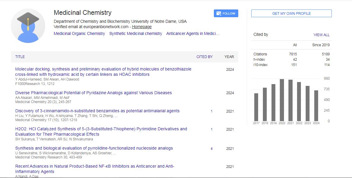
Medicinal Chemistry received 6627 citations as per Google Scholar report