Review Article - (2024) Volume 13, Issue 5
Published: 23-Aug-2021
Introduction: The increased number of documented human coccidian infections that are often indistinguishable from other forms of community-acquired diarrhoea, together with the possibility of treating some of them, suggests a need for proper diagnostic techniques to recover and identify the organism.
Materials and methods: 100 samples which were received in sterile wide mouth container were included in the study. The samples were subjected to macroscopic and microscopic examination by saline mount, iodine mount, modified acid fast stain and auramine and rhodamine staining.
Results: The prevalence of coccidian parasites in this study was observed to be 6%. Coccidian parasites reported were Cryptosporidia and Cystoisospora. Immunocompromised patients were observed to be susceptible to infection with coccidian parasites. Auramine rhodamine stain showed 100% agreement with modified acid fast stain.
Conclusion: This study concluded that Auramine stain is a better and more rapid stain than modified acid-fast stain for identifying coccidian parasites.
Cryptosporidium • Cystoisospora • Immunosuppression• Microscopy
The increased number of documented human coccidian infections that are often indistinguishable from other forms of communityacquired diarrhoea, together with the possibility of treating some of them, suggests a need for proper diagnostic techniques to recover and identify the organism [1]. Human intestinal coccidia includes Cryptosporidium parvum, C. cayetanensis, Isospora belli and Sarcocystis spp. They significantly cause gastrointestinal symptoms [1].
Earlier, Cryptosporidium and Cystoisospora, were assumed to be the causal agents of acute diarrhoea in animals, but recently has emerged as one of the leading causes of prolonged life threatening diarrhoea in immunocompromised patients particularly in patients with AIDS [2].
Coccidian parasites are found to infect intestinal epithelial cells predominantly. They are intracellular cyst forming belonging to apicomplexa protozoa. Mostly transmitted by the fecal-oral route or via contaminated water or food, although Sarcocystis spp is transmitted by improperly cooked meat. Patho physiology involves intestinal inflammation, villus blunting and malabsorption. healthy individuals commonly present with mild to moderate self-limiting diarrhoea during the infective stage, besides asymptomatic infection can also occur. Severe form of the disease is observed in Patients with immune dysfunction who may show severe intestinal injury, prolonged diarrhoea, extreme weight loss, and generalized wasting [1].
Detection of coccidian parasites is mostly through microscopic observation using kinyons acid fast stain [3] this requires use of oil immersion field for screening the smears which is time-consuming and cumbersome. There are other detection methods like antigen assays, IF, PCR which are more sensitive but as they are expensive they are not routinely used in all laboratories. Most of the recommendations in the work-up of community-acquired diarrhoea suggest only bacterial cultures [4]. There is every possibility of missing coccidial infections or delaying the diagnosis. Auramine O (AuO) has widely replaced acid fast stain in mycobacteriology as it is easy and requires less time in screening the smears. However, AuO produces very characteristic images of coccidian oocysts and had yielded good results when used to screen for coccidian parasites [5]. The aim of this study was to investigate if AuO staining is a feasible and inexpensive way to screen for Coccidian parasites in all fecal specimens submitted for O&P examination.
A prospective study done was done at Nizamâ??s Institute of Medical Sciences for a period of 3 months (i.e. from July to September 2019). About 100 fresh stool samples which were received in sterile wide mouth container obtained from immunocompromised patients with history of diarrhoea were included in the study. These samples were subjected to macroscopic examination and microscopy by wet mount, iodine mount, modified acid fast stain and auramine and rhodamine staining. An informed consent was obtained from the patients enrolled in the study. Institutional ethical committee clearance has approved the study.
Method
Fecal samples were collected in wide-mouth disposable containers. Macroscopic examination like colour, consistency and odour were observed. The samples were subjected to a direct wet saline smear and iodine smear.
Direct wet mount
Saline and iodine mount: A drop of saline and Lugolâ??s iodine were placed on left and right halves of the slide and small amount of faeces was mixed by stick to form a uniform suspension. cover slip was placed on the mount and examined under low power objective (10 x) for detection of helminths eggs and larvae; followed by (40x) for protozoan cysts and trophozoites.
Modified acid fast stain
Cold method a thin smear of faeces was made and then fixed with absolute methanol for 1 min. then the slide was flooded with carbol fuschin for 5 mints and rinsed with 50% ethanol for 3-5 seconds and then rinsed with water, followed by decolourization with 1% sulphuric acid for 2 mints or until no colour runs from slide. then the slide was rinsed with water and counter stained with methylene blue for 1min.
Interpretation acid fast oocyst of cryptosporidium and cystoisospora stain pink-red and background stain blue.
Auromine-o stain
A thin smear was prepared on the slide and was heat fixed or methanol fixed for 1 mint. The slide was allowed to cool and was flooded with auromine-o for 15 mints. slide was rinsed with water and flooded with 0.5% acid alcohol for 2 mint and counterstained with potassium permanganate for again 2 mint. slide was rinsed with water, air dried and observed under fluorescent microscope.
Interpretation cryptosporidium and cystoisospora oocysts fluorescence bright and have regular starry sky appearance.
About100 samples were included in the study. The predominant age group was 31 â?? 40 (table 1).
A male preponderance was observed (77) while females were 33. The smear positivity rate was 7% among which the coccidian parasites were predominant (85%) with cryptosporidium and cystoisopora being the commonest and strongyloides larva was observed in one sample.Fig.1. The prevalence of coccidian parasites in this study was 6%. Acid fast stain detected coccidian parasites in 6 samples and the coccidian parasites reported were cryptosporidia and cystoisospora. All patients positive for coccidian parasites were immunosuppressed. The coccidian parasites reported by acid fast stain from 6 samples were also detected by Auramine rhodamine stain which accounted for a 100% agreement of auramine stain with modified acid fast stain.

Figure 1. Parasites detected from stool samples of immunocompromised patients.

Cryptosporidia oocyst
On Modified acid-fast stain Cryptosporidia spp oocysts were observed to be pink-red in colour 4-6 ÃÂ?¼m in diameter with four sporozoites appreciated internally. The background stained uniformly blue.
With Auramine stain, the Cryptosporidium spp cyst showed yellowish-green fluorescence against a dark background and were easily differentiated from other fecal contents by their uniform small size and morphology (Figure 2).

Figure 2. Legend
Isospora oocyst
On Modified acid fast stain the oocysts of Isospora were observed to be ellipsoidal in shape around 23-33 ÃÂ?¼m ÃÂ?? 12-15 ÃÂ?¼m in size with a pink stain. The sporoblast and oocyst wall were also stained. With Auramine stain they showed intense bright yellow fluorescence. (Figure 3). These oocysts were also observed in direct wet mount. The preparation time of Kinyounâ??s acid-fast stain in our laboratory was about 20 minutes and required an additional 5 minutes for screening the slide. The auramine- stain preparation required 17 minutes, while Screening and Interpretation of the fluorescent smears required less than a minute per slide.

Figure 3. Legend
Intestinal coccidian parasites cause disease predominantly in immunodeficient patients, quite a few of them are reported in immunocompetent patients. These can be acquired easily and are difficult to treat. There are battery of tests ranging from microscopy to molecular methods available for detecting these protozoa. But many of these are cumbersome, time consuming thus posing a diagnostic challenge. Therefore, there is need for highly sensitive rapid techniques which aid in early diagnosis and accurate treatment. Though more sensitive methods like antigen detection, nucleic acid amplification assays are available but as they are expensive most of the laboratories still rely on microscopic examination. Kinyounâ??s acid fast stain is used in detection and differentiation of coccidian parasites based on their size and morphology . Auramine stain is a fluorescent dye used in mycobacteriology in detection of acid-fast microorganisms and has replaced Kinyounâ??s acid fast stain because of the ease and sensitivity of interpretation. In India every RNTCP lab is provided with a LED microscope which can be utilised for screening of coccidian parasites without allocation of additional budget. By both techniques, Cryptosporidium spp (3%) and Cystoisospora spp (3%) were the coccidian parasites detected. Similar results were obtained by Abou El-Naga and colleagues, 1998, and Hanscheid and colleagues, 2008. This fluorescent stain could easily differentiate the artifacts from the coccidial parasites, thus yielding better results than Kinyounâ??s acid-fast stain. The advantage of the auramine over Kinyounâ??s acid-fast stain was reported by Abou El-Naga and colleagues in 1998, and by Hanscheid and colleagues in 2008.
The ease of interpretation varied with 2 methods. The screening of Kinyounâ??s acid-fast stained smears were tedious and required more time in interpretation due to examination of fields under oil immersion. And Misdiagnosis due to artifacts was also possible which are commonly seen in fecal samples. The auramine stain on the other hand required much less time as it was extremely easy to visualise fluorescent organisms at ÃÂ??100, with confirmation at ÃÂ??400 magnification. However, the morphological characterization required ÃÂ??1000 oil immersion magnification. Artifacts were easily recognizable by their irregular shape and homogenous staining without welldefined internal structures and hence minimised the misdiagnosis. The ease of this test was noted by Kehl and colleagues in 1995, Ash and Orihel in 1997 and by Mansfield and Gajadhar in 2004 all of whom concluded that the difficulty of interpreting Kinyounâ??s acid-fast stain smears made the fluorescent stain a more desirable test . The reagent cost of the Kinyounâ??s acid-fast stain was less than the auramine stain. Also, the auramine required a fluorescent microscope. MacPherson and McQueen (1993), and Kehl and colleagues (1995 also observed that the cost of fluorescent staining and the availability of the fluorescent microscope are major obstacles to the use of the auramine stain In the present study, Auramine staining had 100% agreement with modified acid fast stain, which is in accordance with earlier studies and may be attributed to the fact that it stains the outer wall of the oocyst as well as internal structures and the phenol present accelerates Auramine penetration through oocyst walls. Cryptosporidium oocysts stain against a dark background and the smears can be easily examined under 20X or 40X objective.Thus Auramine-o stain is better than Ziehl-Neelson stain due to; lower screening time per smear (30 secs vs. 7 min) and its feasibility of screening at low magnification (ÃÂ??400).
We conclude that though modified Ziehl-Neelsen staining technique is considered the gold standard for the detection of coccidian parasites Auramine fluorescent stain should be considered as the screening technique of first choice. This technique is rapid, effortless and does not require much expertise. However, in India detection is restricted to major research laboratories and does not form a part of routine investigations in clinical laboratories; every possibility of missing and delaying diagnosis. therefore, health care providers should precisely request testing for these parasites.
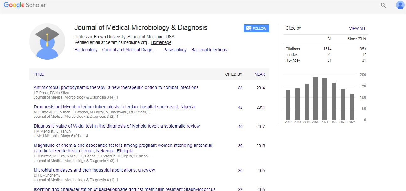
Medical Microbiology & Diagnosis received 14 citations as per Google Scholar report