Mini Review - (2024) Volume 14, Issue 4
Received: 27-Jul-2024, Manuscript No. JBL-24-143504 ;
Editor assigned: 30-Jul-2024, Pre QC No. JBL-24-143504 (PQ);
Reviewed: 13-Aug-2024, QC No. JBL-24-143504 ;
Revised: 20-Aug-2024, Manuscript No. JBL-24-143504 (R);
Published:
27-Jul-2024
, DOI: 10.37421/2165-7831.2024.14.333
Citation: Zhao, Xiaoxian, Feng Lin and Eric D. Hsi. "CD6
as a Therapeutic Target for Autoimmune Disease and Cancer." J Blood
Lymph 14(2024): 333
Copyright: © 2024 Zhao X, et al. This is an open-access article distributed under the terms of the creative commons attribution license which permits unrestricted use, distribution and reproduction in any medium, provided the original author and source are credited.
CD6 is one of the first discovered lymphocyte receptors. It is expressed on all T cells, a NK cell subset, and B-lymphocyte B1a subsets. CD6 is involved in cell-to-cell interaction and modulation of adaptive immune responses. It is also implicated in the pathogenesis of a variety of immune-mediated conditions. CD6 and its two major known ligands (CD166 and CD318) have emerged as new therapeutic targets for autoimmune diseases. Recent studies explored the distribution of CD6 in both T-cell lymphomas and aggressive NK/T cell neoplasms, and evaluated the activities of CD6-antibody drug conjugate in both in vitro and in vivo mouse models for these malignancies. In this mini-review, we highlight the current understanding of CD6 as a therapeutic target in autoimmune disease, T cell lymphoma and aggressive NK/T cell neoplasms.
CD6 • T-cell lymphoma • Extranodal NK-cell lymphoma • Aggressive NK-cell leukemia • Antibody-drug conjugate
CD6 is a 105-130 kD type I transmembrane glycoprotein member of the Scavenger Receptor Cysteine-Rich Superfamily (SRCR-SF). It contains an extracellular component with three SRCR domains and an intracellular domain with multiple phosphorylation sites that modulate recruitment of signal transduction proteins. CD6 is constitutively expressed on T-cells, some NK cell subsets and 1% of B cells caller B1a cells. There are two major known ligands for CD6, CD166 and CD318. CD166 (ALCAM) Activated Leukocyte Cell Adhesion Molecule is expressed on activated T-cells, monocytes, endothelial cells, epithelial cells and synovial fibroblasts. CD318 (CDCP1, CUB Domain Containing Protein 1) is expressed on most epithelial cells, some stem cells and many solid tumors [1]. CD6 has been primarily thought to function as a costimulatory molecule and adhesion molecule since interaction with ALCAM on Antigen Presenting Cells (APCs) stabilizes the T-cell-APC interaction, supporting cell proliferation and differentiation to Th1 and Th17 cells [1]. Blocking antibodies to CD6 inhibit contact induced T-cell proliferation and dual crosslinking of CD3 and CD6 results in proliferation similar to CD3/CD28 co-crosslinking [2]. Recent studies demonstrate a signaling function for the intracytoplasmic domain, with phosphorylation sites mediating potential interactions with tyrosine and serine kinases and phosphatases, and/or signaling adaptors. Mutagenesis studies eliminating phosphorylation-target residues in the intracytoplasmic domain of CD6 resulted in increased activation of T-cells, compatible with loss of inhibitory signaling function [3].
Interest in manipulating CD6 signaling stems from the initial paradigm of CD6 acting to enhance T-cell activation. Interfering with CD6-ALCAM and CD6-CD318 interactions are thus of interest in autoimmune disease [1]. For example, CD6 knockout mice are protected from experimental autoimmune encephalitis and collagen induced arthritis which serve as models of Multiple Sclerosis (MS) and Rheumatoid Arthritis (RA), respectively. Indeed, treatment with antibodies to CD6 in CD6 humanized mice attenuated diseases in both of these preclinical models [4,5]. Other diseases such as Sjogren syndrome and inflammatory bowel disease are also areas of interest for CD6 antibody treatment [1]. Psoriasis has been a major area of interest, with CD6 knockout animal models and clinical evidence supporting a role for CD6 in the treatment of this disease through inhibiting Th17 polarization [6]. Preclinical and clinical investigations of itolizumab, a humanized CD6 antibody, have resulted in its approval in India for the treatment of chronic plaque psoriasis [7].
The field of immuno-oncology has rapidly developed based on the realization that the Tumor Microenvironment (TME) and immune system plays a key role in cancer progression. This is supported by the understanding of the importance of immunosuppressive factors such as Transforming Growth Factor Beta (TGF-β), suppressive cells such as Tregs and immune checkpoint pathways in the TME. With regard to CD6, experimental evidence now suggests that UMCD6, a Mouse anti-human-CD6 monoclonal antibody, can activate cytotoxic T-cells and NK cells to enhance tumor cell killing in experimental in vitro and in vivo systems [1,8]. Given these data and the fact that CD6 may also act as a target in lymphoid malignancies that naturally express CD6, we explored expression patterns and effects in more detail in T and NK cell neoplasms.
T- and NK-cell lymphomas are a heterogeneous group of hematological malignancies which includes more than 30 subtypes in the WHO classification of hematolymphoid tumors, 5th edition [9] T-Cell Lymphomas (TCLs) are broadly classified into systemic and cutaneous types based on their clinical features. The most common systemic subtypes include Peripheral TCL Not Otherwise Specified (PTCL-NOS), Angioimmunoblastic TCL (AITL) and Anaplastic Large Cell Lymphoma (ALCL), ALK Positive and Negative (ALK ±). Most systemic subtypes of TCL have an approximate 5-year Overall Survival (OS) of ≤ 35% except for ALCL, ALK+ (80%) and ALCL, ALK- (50%) [9]. Frequent Cutaneous TCL (CTCL) subtypes include mycosis fungoides/sezary syndrome and CD30-positive lymphoproliferative disorders. Natural Killer (NK) cell lymphomas and leukemias are rare in North America and Europe but seen more frequently in Asia. Aggressive NK-Cell Leukemia/Lymphoma (ANKLL) is an uncommon, highly lethal malignancy derived from NK cells. No FDA-approved therapy exists for this disease. Most cases are resistant to therapy and the median survival time is measured in months [9]. Extranodal NK/T-cell Lymphoma (ENKTL) is a rare subtype of non-Hodgkin lymphoma, a sizeable minority of ENKTL patients present with advanced stage disease and have a poor prognosis [10]. Like ANKLL, and there is no standard therapeutic regimen for ENKTL. Development of model systems and effective targeted therapeutic agents for T and NK cell malignancies remains an unmet need.
Antibody-Drug Conjugates (ADCs) are combination of a monoclonal Antibody (mAb) with a cytotoxic molecule payload connected via a chemical linker. They are developed to target tumor cells via the antibody specificity and mediate selective cytotoxicity. In lymphoma therapy, the success of brentuximab vedotin, an ADC targeting CD30 in ALCL, Hodgkin lymphoma and other CD30+ TCLs provides the impetus to explore additional targets in T and NK-cell malignancy [11,12]. Given the expression pattern of CD6 in normal T and NK cells and its demonstrated role in modulating T-cell response in autoimmune disease, we believe exploration of CD6 as a therapeutic target in lymphoma is warranted.
The expression pattern of CD6 in TCL cell lines, Patient-Derived Xenograft (PDX) TCL models and primary human TCLs was reported in 2023 [13]. Cell lines representing CTCL (HH and Hut78), T-lymphoblastic leukemia (Jurkat) and selected 6 PDX models (three AITL, one PTCL-NOS, one CTCL and one NK/TCL) were analyzed for CD6 expression by flow cytometry and all samples were positive for CD6. Immunohistochemistry (IHC) staining of 59 TCLs from a Tissue Microarray (TMA) was performed (34 PTCL-NOS, 17 AITL, 4 ALCL, one intestinal T-cell lymphoma, one post-transplant TCL and two NK/T cell lymphomas). CD6 positive cases were defined as membranous staining in ≥ 10% of tumor cells and CD6 was scored with positive percentage of tumor cells and staining intensity. 90% (53/59) cases expressed CD6. Recent studies in aggressive NK cell neoplasms also showed that CD6 was expressed in the majority of tested cases (68.3%, N=41) by IHC [14]. These staining results were summarized in Table 1 and illustrated in Figure 1. Given that CD6 is expressed in most T and NK cell lymphomas, further investigation in functional models was warranted.
| Subtype of tested NHLs | Case number | % of CD6+ | % of cases with >50% tumor cells CD6+ |
|---|---|---|---|
| PTCL-NOS | 34 | 100 (34/34) | 82.4 (28/34) |
| AITL | 17 | 94.1 (16/17) | 76.5 (13/17) |
| ALCL | 4 | 25 (1/4) | 0 (0/4) |
| Intestinal T-cell lymphoma | 1 | 100 (1/1) | 100 (1/1) |
| NK/T cell lymphoma | 2 | 50 (1/2) | 0 (0/2) |
| Post-transplant TCL | 1 | 0 (0/1) | 0 (0/1) |
| ANKLL | 10 | 70 (7/10) | 50 (5/10) |
| ENKTL | 31 | 67.7 (21/31) | 54.8 (17/31) |
UMCD6 is efficiently internalized by TCL cells upon binding to CD6 on the surface of malignant T cells [13]. Functional analysis of a CD6-ADC (UMCD6 conjugated with Monomethyl Auristatin E, MMAE) showed in vitro cytotoxic activity against both TCL and ANKL cell line in a dose and time-dependent manner. In addition, in vivo studies using an HH cell line xenograft in NOD-scid IL2R gamma null (NSG) mice demonstrated that treatment with CD6-ADC could both prevent the development of tumors and effectively diminished tumor sizes at later stages. Local administration of low-dose of CD6-ADC also had anti-tumor activity. A humanized UMCD6 has been produced with a binding affinity comparable to the parental UMCD6 and effectively reduced established TCL tumor and metastasis in xenografted NSG mice [13]. In an ANKL-PDX study, a single dose of murine UMCD6-ADC blocked the engraftment of tumor cells (Figure 1) [14].

Figure 1. CD6 IHC in a representative NK/T-cell lymphoma (Left panel 40x, right panel 400x). Staining was performed on the Ventana DISC ultra-platform with a monoclonal (ERP4057) CD6 antibody.
These data support further investigation of CD6 as a potential novel therapeutic target for TCL and aggressive NK/T cell neoplasms. The CD6-ADC (MMAE) has the advantages of 1) high selectively for targeted lymphocytes only and 2) preferential effect on proliferating tumor cells by mitotic toxin MMAE, potentially limiting toxicity in normal CD6+ T and NK cells. In the pre-clinical mouse study the CD6-ADC prevented the engraftment of tumors, effectively diminished tumor sizes at later stages, and showed anti-tumor efficacy with low-dose intratumoral injections [13]. Furthermore, CD6-ADC also strongly prevented the engraftment of ANKL tumor cells [14]. It should be noted that approximately 10% of TCL cases and 30% of tested ANKLL/ENKLL cases were CD6 negative by IHC in formalin fixed paraffin embedded archival tissues, and the percentage of CD6+ tumor cells were different among the cases (10%-100%). Such variability in CD6 expression may reflect the heterogeneity and complexity of these malignancies or could also be a technical or sensitivity issue since archival tissue IHC studies may be less sensitive that other modalities such as flow cytometry in fresh samples.
CD6 is expressed on T-cells and a subset of NK cells and plays a role in modulating T-cell activation. Targeting CD6 is a therapeutic strategy in autoimmune disease and, more recently T and NK cell malignancies. With regard to the latter, preclinical studies support the rational for further clinical development of the humanized CD6-ADC as a targeted agent in these aggressive lymphoid malignancies.
[Crossref] [Google Scholar] [Pubmed]
[Crossref] [Google Scholar] [Pubmed]
[Crossref] [Google Scholar] [Pubmed]
[Crossref] [Google Scholar] [Pubmed]
[Crossref] [Google Scholar] [Pubmed]
[Crossref] [Google Scholar] [Pubmed]
[Crossref] [Google Scholar] [Pubmed]
[Crossref] [Google Scholar] [Pubmed]
[Crossref] [Google Scholar] [Pubmed]
[Crossref] [Google Scholar] [Pubmed]
[Crossref] [Google Scholar] [Pubmed]
[Crossref] [Google Scholar] [Pubmed]
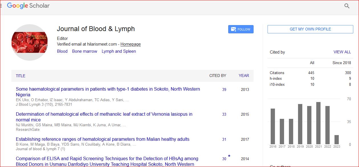
Journal of Blood & Lymph received 443 citations as per Google Scholar report