Commentary - (2024) Volume 14, Issue 4
Received: 07-Oct-2024, Manuscript No. JBL-24-149688 ;
Editor assigned: 09-Oct-2024, Pre QC No. JBL-24-149688 (PQ);
Reviewed: 22-Oct-2024, QC No. JBL-24-149688 ;
Revised: 28-Oct-2024, Manuscript No. JBL-24-149688 (R);
Published:
06-Nov-2024
, DOI: 10.37421/2165-7831.2024.14.332
Citation: Mei, Yihan and Runxia Gu. ''Commentary: A New Marker for Tumor-Reactive T Cells in Acute Myeloid Leukemia." J Blood Lymph 14(2024): 332
Copyright: © 2024 Mei Y, et al. This is an open-access article distributed under the terms of the creative commons attribution license which permits unrestricted use, distribution and reproduction in any medium, provided the original author and source are credited.
The field of cancer immunotherapy has witnessed remarkable advancements over the past decade, transforming the landscape of cancer treatment. Our recent publication in experimental hematology and oncology provides critical insights into the specific profile of tumor-reactive T cells in the bone marrow of Acute Myeloid Leukemia (AML) [1]. This commentary aims to review the key findings of the study and discuss their implications for future research and clinical applications.
In brief, our study reveals that the AML tumor-reactive T cells exhibited non-exhausted but cytotoxic and senescent-like features with upregulated Natural Killers (NK) markers. In addition, ADGRG1 was specifically upregulated in AML tumor-reactive T cells. The ADGRG1+CD8+T cells from AML patients showed significantly higher IFN-γ releasing level and cell-killing ability. AML patients with a lower proportion of ADGRG1+CD8+T cells at diagnosis showed relatively poor survival outcomes. These results indicated the potential role of ADGRG1 in identifying and enriching tumor-reactive TCR sequences.
The profile of tumor-reactive T cells in solid tumors has been well studied and they are presented as terminally exhausted T cells with upregulated markers like PD-1 [2]. However, our research group found that tumor-reactive T cells showed much different phenotype in AML. This is partly due to the disseminated nature of AML blasts which are aggressively progressed and systemically distributed in the loosely structured Bone Marrow (BM) and Peripheral Blood (PB). In this low-antigen niche, T cells tend to differentiate into a killer cell lectin-like receptor-expressing cytotoxic phenotype [3].
While TCR-T showed significant treatment efficacy in specific cancer types [4], the TCR-T construction is difficult. One important reason is that the steps of traditional TCR-T construction workflow are indeed time-consuming, including 1) neoantigen prediction; 2) experimental enrichment of presented neoantigen; 3) screening of T cells recognizing neoantigens and 4) construction of TCR-engineered T cells. After the identification of ADGRG1, we could develop a machine-learning model based on ADGRG1-enriched tumor-reactive T cells, to predict and construct personalized TCR-T therapies. In this way, we can bypass the complexity of MHC typing and neoantigen prediction, which significantly simplifies the workload and increases the accessibility of TCR-T for clinical therapy applications. A recent study constructed a machine learning model predicTCR based on CXCL13-identified Tumor-Infiltrating T cells (TILs) to detect TILs across different solid tumor types, which also demonstrates the feasibility of the potential clinical usage of ADGRG1 in AML [5].
In conclusion, the evolving landscape of cancer immunotherapy, particularly in the context of TCR-engineered T-cell therapies, presents promising avenues for enhancing treatment efficacy in acute myeloid leukemia. Our study not only enriches the understanding of T cell dynamics in AML but also paves the way for innovative, personalized therapeutic approaches.
[Crossref] [Google Scholar] [Pubmed]
[Crossref] [Google Scholar] [Pubmed]
[Crossref] [Google Scholar] [Pubmed]
[Crossref] [Google Scholar] [Pubmed]
[Crossref] [Google Scholar] [Pubmed]
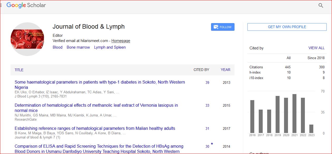
Journal of Blood & Lymph received 443 citations as per Google Scholar report