Review Article - (2020) Volume 4, Issue 2
Received: 10-Jul-2020
Published:
03-Aug-2020
, DOI: 10.37421/jcao.2020.4.120
Citation: Nabil A Shallik, Nissar Shaikh, A Raju Vegesna, Ahmed Lafir Aliyar and Yasser Ali Hammad. "COVID-19 Pandemic and Embolic Manifestations; Old Topic and a New Visit". J Clin Anesthesiol 4 (2020) doi: 10.37421/JCAO.2020.4.120
Copyright: © 2020 Shallik NA, et al. This is an open-access article distributed under the terms of the Creative Commons Attribution License, which permits
unrestricted use, distribution, and reproduction in any medium, provided the original author and source are credited.
Perioperative embolism increases the risk of morbidity and mortality in surgical patients. Pulmonary Embolism (PE), Fat Embolism Syndrome (FES), and Vascular Air Embolism (VAE) are a relatively common embolic phenomenon in the perioperative period. Surgical intervention causes tissue injury, hypercoagulability, and venous stasis. The incidence of pulmonary embolism varies with the type of surgical interventions, and hip hemi-arthroplasty has a higher incidence, whereas the laparoscopic surgeries have a lower incidence of pulmonary embolism. Various risk predispose to a perioperative pulmonary embolism. CTPA (Computerized Tomographic Pulmonary Angiography) has high sensitivity and specificity for the diagnosis of pulmonary embolism. Unfractionated Heparin (UFH) should be started as soon as pulmonary embolism is suspected. FES is the organ dysfunction caused by fat emboli. FES can be diagnosed by using a combination of clinical criteria and imaging studies. Supportive care is the mainstay of treatment for FES while heparin, steroid, and dextran are not recommended. VAE is frequent in obstetric, laparoscopic, and neurosurgical surgeries. VAE is increasingly occurring in divers, aviators, and astronauts due to the dysbarism. VAE commonly manifests by respiratory, cardiogenic, and neurological manifestations. Treatment includes hyperbaric oxygen therapy, UFH, and lignocaine. The incidence of pulmonary embolism in ICU patients with COVID-19 range between (14-43)%, most of them on anticoagulants. The diagnosis is challenging. The raised D-Dimer is an indication to do CTPA (Computerized Tomographic Pulmonary Angiography).
Fat embolism • Heparin • Pulmonary embolism • Surgery • Vascular air embolism
Various types of embolisms can occur during the perioperative period, namely thromboembolism, fat, septic, amniotic, tumor, gestational trophoblastic, and air embolisms. The more common emboli occurring during this period are thrombus, fat, and air embolisms. These embolisms lead to a grave prognosis with increased morbidity and mortality [1]. The exact etiology and impact of these emboli are unknown as the milder cases go unnoticed, and only moderate to severe thromboembolism is diagnosed. Despite the improvement in technologies, the diagnosis and management of perioperative thromboembolism remains a challenge [1].
Perioperative pulmonary thromboembolism
Anesthetists may find themselves responsible for the diagnosis and management of perioperative thromboembolism, and if diagnosed intraoperatively the ongoing surgical interventions will interfere with the management [1].
Increased incidence: Many folds are growing in the occurrence of postoperative thromboembolism as the surgical trauma, venous stasis, and hypercoagulability fulfill the Virchow’s triad of thrombosis. The interesting fact is that 50% of patients developing postoperative thromboembolism were on Deep Venous Thrombo-Prophylaxis (DVT) [2].
The incidence of pulmonary embolism differs according to the type of surgery (Table 1). The perioperative pulmonary embolism commonly occurs (up to 30%) after the orthopedic surgery. Among these surgeries, hip arthroplasty is frequent to cause pulmonary embolisms (up to 24%), as the location of surgery will have an impact not only on venous return but also a distortion of the femoral vein. The laparoscopic surgeries have a lower incidence of pulmonary thromboembolism due to lesser tissue trauma and early mobilization [3].
| Surgical population | Incidences of PE |
|---|---|
| General surgery | 1.6% (average) |
| Thoracic | 1.5%-2% |
| Abdominal | 0.32%-1.0% |
| Laparoscopic | 0.06%-0.9% |
| Vascular | 0.4%-0.7% |
| Head and neck | 0.4%-0.44% |
| Gynecologic | 0.3%-4.1% |
| Ortho | 0.7%-30% |
| Ortho: hip fracture | 4.3%-24% |
| Urologic | 0.9%-1.1% |
| Neurosurgical | 0%-4% |
| Trauma | 2.3%-6.2% |
| Acute SCI (Spinal Injuries) | 4.6%-9% |
Diagnosis: It is relatively easier to diagnose pre and post-operative pulmonary embolism by the signs and symptoms associated with pulmonary embolism and by using various clinical criteria. Whereas, the determination of intraoperative pulmonary embolism is challenging, as desaturation, decrease in the End- Tidal CO2 (ETCO2), increased airway pressure, electrocardiographic changes, and hypotension are not specific findings. When the decrease in ETCO2 is associated with a simultaneous increase in the arterial PCO2, it is suggestive of acute pulmonary embolism during the intraoperative period, and it is called as separation phenomenon [4].
The echocardiography will can be a great of help in the intraoperative period, it may show typical McConnell sign (Thinning of right ventricular wall with apical sparing and hyperkinesia) or systolic flattening of interventricular septum with right ventricle dilatation and tricuspid regurgitation [5].
The better way to confirm the PE is CTPA (Computerized Tomographic Pulmonary Angiography) as it is specific and sensitive (more than 95%) for the diagnosis of PE. CTPA is available 24 × 7 in most hospitals and can give alternative diagnoses in the absence of the PE [5]. The disadvantage of CTPA is that it gets abused. The V/Q (Ventilation/Perfusion) scan is not useful as it is not conclusive in 50% of the suspected PE cases. V/Q SPECT (Single Photon Emission Tomographic V/Q) scan is also highly sensitive for diagnosing PE, but it gives binary interpretation (PE present or absent). The conventional catheter pulmonary angiography has become a historical gold standard for the diagnosis of PE [5].
Pathophysiology: The European Society of Cardiology described perioperative pulmonary embolism as a spiral of hemodynamic collapse. The PE causes RV (Right Ventricle) dilatation, leading to tricuspid regurgitation, increased RV wall tension, increased oxygen demand, septal shift, decreased cardiac output and obstructive shock [5].
Risk factors: The risk of PE increases in elders, obese, bedbound, carcinoma patients, and patients with inflammatory disease. The orthopedic, thoracicabdominal, and vascular surgeries pose a risk for perioperative pulmonary embolism: general anesthesia and failure to administrate the DVT prophylaxis cause higher perioperative PE. The hereditary deficiency of natural anticoagulants will also increase the risk of the occurrence of pulmonary embolisms [6]. Apart from the group mentioned above, patients with Arterial Fibrillation (AF), Myocardial Infarction (MI), ischemic stroke, and diabetes mellitus are also at the higher risk of perioperative pulmonary embolism [7].
Treatment of PE: In suspected cases of PE, anticoagulation with unfractionated heparin should be started immediately. When signs of right ventricular failure are present, and the patient is dehydrated fluid boluses will be useful, noradrenaline helps by inotropic and increasing the blood pressure; dobutamine should be used along with norepinephrine and not as a sole agent, to avoid hypotension and arrhythmias [5].
Fulminant pulmonary embolism: Up to 41% of patients with massive (Fulminant) pulmonary embolisms will present as cardiac arrest. In these patients, along with the immediate CPR (Cardiopulmonary Resuscitation), intravenous thrombolysis is essential. Due to its availability, rapid administration, and faster action, tPA (tissue Plasminogen Activator is the drug of choice in a dose of 0.6 mg/Kg over 15 minutes (maximum of 50 mg) and 100 mg over 2 hours [5,8].
COVID-19 and pulmonary embolism
A novel coronavirus (SARS CoV-2) surfaced in China towards the end of 2019, producing acute respiratory distress in affected individuals resulting in COVID-19 (Coronavirus Disease 2019). The WHO declared COVID-19 a pandemic on March 11th, 2020.
Initial management guidelines were based on the assumption that COVID 19 patients developed ARDS (Acute Respiratory Distress Syndrome). As our understanding of the disease expands, there is emerging evidence that COVID-19 is associated with vascular dysfunction, hypercoagulability, and thrombosis.
Recent evidence suggests that pulmonary embolism in ICU patients with COVID-19 can range between (14-43)% [9-13]. Most of these patients were either on prophylactic or therapeutic anticoagulation. Moreover, some had additional risk factors like cancer, recent surgery, and a history of VTE. Data for non-ICU patients and outpatients is lacking. Some autopsy studies have demonstrated microvascular thrombosis in the lungs [14,15].
Pathogenesis: Although it is not possible to attribute the increased incidence of pulmonary embolism in COVID-19 patients to be the specific effects of SARS CoV-2, the process of epitheliopathy and hypercoagulability may be due to an interplay between hypoxia, exaggerated immune response, and thrombo-inflammation.
The pathogenesis of hypercoagulability in COVID-19 patients is incompletely understood, as the endothelial injury occurs due to a multitude of factors; direct injury by the virus [13]
• Cytokine mediated (IL-6)
• Complement mediated
• Intravascular catheters
Patients with COVID-19 have been associated with several coagulation abnormalities that can be attributed to hypercoagulability;
• Elevated factor VIII
• Elevated fibrinogen
• Elevated levels of D-Dimer
• Antiphospholipid antibodies
• Increased factor VIII antibodies
SARS CoV-2 invades human cells by binding to surface ACE-2 receptors, including endothelial cells [7]. ACE-2 is essential for the metabolism of Angiotensin II to the peptide angiotensin [9-15]. Angiotensin II is a potent vasoconstrictor, endothelial activator, and pro-inflammatory. Whereas, angiotensin [9-15] has anti-inflammatory and vasodilatory properties. In COVID-19 patients, ACE-2 consumption occurs due to the internalization of the SARS CoV-2/ACE-2 complexes. Consequently, a state of imbalance between angiotensin II and angiotensin [9-15] leading to a thrombotic state. Admission levels of angiotensin II are twice normal in COVID-19 patients [16].
Several factors indicate that the endothelium plays a pivotal role in the pathogenesis of hypercoagulability. The hypoxia seen in COVID-19 patients results in vasoconstriction and reduced blood flow, leading to endothelial dysfunction [17,18]. Moreover, COVID-19 induced pro-inflammatory cytokines to result in endothelial injury that causes the release of Ultra-Large von- Willebrand Factor (ULvWF) multimers and tissue factor [17,19-21].
Critically ill COVID-19 patients exhibit a platelet/ULvWF complex attached to the injured pulmonary endothelium and intra-alveolar fibrin deposition, forming microthrombi [17,22].
Ultimately, low blood flow due to stasis and vasoconstriction, endothelial injury, and hypercoagulability (Virchow’s triad) puts severely ill COVID-19 patients at higher risk thrombosis.
Evaluation and management: Current literature indicates that the features of COVID-19 patients with respiratory distress differ from those seen with classical ARDS. COVID-19 patients develop profound hypoxemia early in the disease, which is not associated with overt respiratory dysfunction. Experience with intubated patients suggests that; there is only a mild to moderate reduction in their pulmonary compliance. However, later in the course of the disease, they develop more consistent features with ARDS. This presentation alludes to the possibility of an alternate underlying mechanism like pulmonary vascular dysfunction and thrombosis.
Evaluation and management of these patients are challenging due to the lack of evidence and consensus on the subject. The International Society has published interim guidance on Thrombosis and Hemostasis (ISTH), and frequently asked questions appear on the websites of the American Society of Hematology (ASH) and the American College of Cardiology (ACC).
Diagnosis of PE-a normal D-Dimer is enough to exclude PE. However, a raised D-Dimer is not specific for PE. If D-Dimer rise and other factors suggesting PE like hypoxia, hemodynamic instability, and VTE are present, CTPA is the preferred test. However, performing a CTPA in unstable COVID-19 patients can be challenging. Moreover, all infection control measures should be strictly followed, making imaging in COVID-19 patients more challenging. V/Q scan is an alternative if CTPA cannot be performed or is inconclusive. It can be challenging to interpret V/Q scan results in COVID-19 patients with significant underlying pulmonary disease.
Fat embolism syndrome
Long bone fractures produce fat emboli, and when these emboli cause organ dysfunction particularly the lung, brain and skin this triad of (lung, brain and skin dysfunction) is called Fat Embolism Syndrome (FES) [23]. Zenker in 1862 described FES at autopsy and within a decade Von Bergman Clinically diagnosed FES in fracture femur patient [23].
Epidemiology: The incidence of FES varies from 1% to 29%, the exciting fact is that in the retrospective studies reported an incidence of<1, if diagnosed based on clinical criteria the incidence of FES was 0.9%, whereas in the autopsy the incidence was 20% and in the prospective study the FES was reported to be (11-29)% [23-25].
Etiology: FES caused by either traumatic or medical etiology. FES is more frequent with the fracture of long bones, rarely with liposuction and soft tissue injuries. The medical reasons for FES are severe pancreatitis, steroid therapy, sickle cell hemoglobinopathies and fat emulsion infusion [23,26].
Risk factors: Young age, closed long bone fracture, conservative therapy for fracture is the risk for development of FES [23]. The risk factors for FES after intramedullary nailing are over-zealous nailing, reaming of the medullary cavity, increased velocity of nailing, increase gap between cortical bone and the nail.
Pathophysiology: There are mainly two theories for the development of FES. Gossling et al., proposed the mechanical theory for the FES, where the large fat droplets are released into the circulation get deposited into the capillary bed and travel to the brain and distal organs through the AV (Arteri-Venous) shunts [27]. Baker et al., proposed the biochemical theory for FES, trauma causes the release of chylomicron into the circulation, the acute phase proteins lead to coalescing of chylomicron, then these chylomicrons are hydrolyzed to fatty acid causing distal organ dysfunctions. The biochemical theory explains the delay of 24-72 hours for the occurrence of FES after the initial insult, this is the time taken to form fatty acid from the fat globules [28].
Diagnosis: FES is the diagnosis of exclusions. The triad of organ dysfunction in a high-risk group (cerebral dysfunction, respiratory impairments, and skin petechiae) typically occurring after 24 to 72 hours of the insult should give a high index of suspicion for the FES. FES patients deteriorate with confusion and oxygen desaturation; Arterial Blood Gas (ABG) will show PaO2 (Partial pressure of Oxygen) of around 60 mmHg, skin rash may be seen in the upper portion of the body particularly around axilla. FES is diagnosed with lower accuracy by using clinical criteria, namely Guard and Wilson, Schonfield’s, and Lindique’s criteria’s, by using Lindique’s criteria, one can diagnose the FES from the respiratory parameters [23].
Imaging studies play a crucial role in confirming the FES; hence the combination of the clinical criteria and imaging studies are increasingly used for the diagnosis of FES [29]. Chest x-ray will show a sandstorm appearance, CT (Computerized Tomography) of the chest will show the typical alveolar consolidation, CT brain may be normal, or it shows edema whereas the MRI (Magnetic Resonance Imaging) will show star studded appearance in the diffusion phase (Figures 1 and 2) [23,29-31].

Figure 1. Chest x-ray showing sandstorm appearance.

Figure 2. CT and MRI brain showing FES changes.
Treatment: The mainstay of treatment for FES remains supportive. The use of steroids, heparin, dextran, and alcohol is not useful and not recommended. FES patients may need invasive ventilation and vasopressors' support. The shock will exacerbate the lung injury; hence optimization of intravascular fluid status is essential. Use of Albumin has double advantages; it minimizes the effect of free fatty acids on organs and maintains the intravascular volume [23].
Prevention: Early fixation, external fixation, venting the medullary cavity, and use of steroids in the preoperative period may reduce the occurrence of FES [32].
Vascular air embolism
Although vascular air embolism has been known since the nineteenth century, the interest and reporting of VAE increased over three decades. VAE is defined as the entrainment of air or gas from the operative field or communication with the environment into the venous or arterial vasculature and producing systemic impact or production of air bubbles in the circulation by dysphoric barotrauma causing VAE in astronauts, aviator and SCUBA (Self- Contained Underwater Breathing Apparatus) divers [33].
Epidemiology: Exact incidence of VAE is not known as the mild cases go unnoticed. VAE is increasing with dysphoric barotrauma. In SCUBA divers, it is the 2nd most common cause of death. Obstetric surgeries, neurosurgeries in sitting position, laparoscopic surgeries are the common cause for the perioperative VAE [34].
Etiology: The perioperative VAE is common. When the surgical site is above the level of heart or the operative field contains lots of uncompressed venous channels. The second etiology for VAE is the creation of the pressure gradient, which facilitates the air entry into the venous system. For instance, insertion or removal of Central Venous Catheter (CVC), the third etiology of VAE is mechanical insufflation or pressure infusions such as GI endoscopies and laparoscopic surgeries. The fourth etiology is barotrauma commonly due to penetrating chest injuries. The fifth Cause of VAE is sudden changes in barometric pressures (dysphoric) barotrauma generating air bubbles in the circulation [33-35].
Risk factors: [33]
• Operative field 5 cm above the levels of heart
• An operative field full of non-compressible venous plexuses
• During insertion of CVC failure to occlude the needle hub, fracture or detachment of catheter, hypovolemia, deep inspiration, and upright position
• SCUBA divers, too rapid ascent, too long diving, diving at depth and cold water, alcohol intake, and older age
Pathophysiology: Smaller air bubbles get absorbed from the circulation and do not cause any clinical manifestation. A moderate amount of air will cause pulmonary vascular injury and leads to pulmonary hypertension and edema. Lager boluses of air (200 to 300) ml will cause airlock of the right heart causing obstructive shock and immediate death [33,35].
In up to 30% of patients, the Foramen Ovale (PFO) will be patent, allowing the air or other emboli from the right side of the heart to enter the left side, called paradoxical embolism causing arterial embolism of the vital organs (Figure 3) [36].

Figure 3. Paradoxical air embolism to brain.
Clinical manifestations: VAE presentations can vary according to the speed of air entrainment, nature, and volume of the air circulation, commonly manifested by respiratory, cardiac, and neurological dysfunctions. Respiratory manifestations include tachycardia, dyspnea, pulmonary edema, and hypoxia. Cardiac manifestations are chest pain, arrhythmias, and PEA (Pulseless Electrical Activity). The neurological manifestation ranges from headache to drowsiness [34,35].
Diagnosis: A high index of suspicion in risky patients is the cornerstone for the diagnosis of VAE. Trans-esophageal echocardiography is most sensitive in detecting VAE, bubbles as small as (5 to 10) microns will be detected.
Precordial Doppler Ultrasound is the most sensitive non-invasive monitor; it can detect as little as 0.05 ml/Kg of the air bubbles in the circulation. End- Tidal Nitrogen (ETN2) will show the changes earlier than the ETCO2 (End-Tidal Carbon Dioxide) [33-35].
The use of hyperbaric oxygen therapy is useful in air embolism as it causes compression of air bubbles, speedy dissolution of bubbles, and improving the oxygenation of ischemic tissues [37,38].
Some case reports suggest that the use of heparin and local anesthetic lignocaine reduces the severity of VAE on various organ dysfunctions [37].
Morbidity and mortality: The outcome of VAE patients depends on the rate of air accumulation and the patient's position at the time of embolism [33,34,37].
Although there is an improvement in the diagnosis and management of VAE, the mortality remains high around 80% [33,34,37].
VAE can be prevented up to some extent by operating in park-bench position instead of sitting position, 50 degree reveres Trendelenberg position reduces the chances of VAE from (44 to 1)% in obstetric patients, optimizing intravascular fluid status, avoiding the use of nitrous oxide and in SCUBA divers using diving tables and follow the algorithm for ascent and descent [39].
Treatment of VAE: [37]
• Prevent further air entry into the circulation by covering or flushing the operative filed with saline
• Reduction in volume of air entrained by the administration of 100% oxygen or hyperbaric oxygen therapy
• Optimization of fluid status and hemodynamic support by vasopressor and inotropes
• Aspiration of air from the right atrium using Bunegin-Albin multi-orifice catheter had a success rate of 60%
• Rapid CPR (Cardio-Pulmonary Resuscitation) will dislodge the air from the right side of the heart and relieve the obstruction and return of spontaneous circulation
The perioperative embolisms occur due to various types of emboli; the common are thromboembolic pulmonary, fat, and vascular air embolisms. The incidence of pulmonary embolism increases by many folds as surgical intervention leads to fulfillment of the Virchow's triad. There are various patients, surgical, and medical risk factors for perioperative pulmonary embolism. Pulmonary embolism causes right ventricular dilatation, septal shift, and obstructive shock. Diagnosis of intraoperative pulmonary embolism is challenging, separation phenomenon and echocardiography may help in diagnosis during this period, and CTPA is confirmatory. As soon as we suspect the pulmonary embolism, patients should be started on UFH. In cases of fulminant pulmonary embolism leading to cardiac arrest, thrombolysis should be initiated.
Fat Embolism Syndrome (FES) occurs commonly after long bone fractures, and the diagnoses are by a combination of clinical criteria and imaging studies. The management of FES is conservative and supportive. FES can be prevented by early fixation of the fractures and methylprednisolone's use in the preoperative period.
Vascular Air Embolism (VAE) is perhaps the most common perioperative embolism, apart from surgery and trauma, aviation and SCUBA diving are the other frequent etiologies for VAE. There are various risk factors for VAE from the operating site above the heart to the rapid ascent and descent during the SCUBA diving. The clinical manifestations vary according to the speed of air entrainment and the position of the patient. The trans-esophageal echo and precordial Doppler detect even small bubbles in the circulation. Hyperbaric oxygen therapy helps to treat VAE patients. The mortality from VAE remains high.
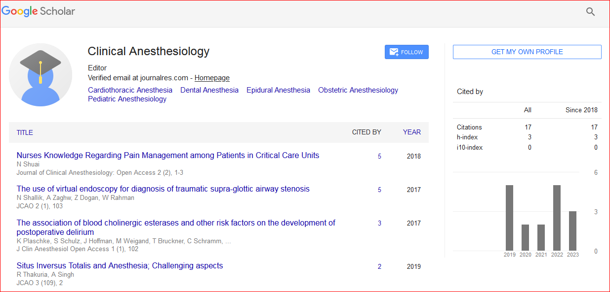
Journal of Clinical Anesthesiology: Open Access received 31 citations as per Google Scholar report