Editorial - (2022) Volume 9, Issue 1
Received: 01-Jan-2022, Manuscript No. bset-21-34113;
Editor assigned: 04-Jan-2022, Pre QC No. P-34113;
Reviewed: 06-Jan-2022, QC No. Q-34113;
Revised: 12-Jan-2022, Manuscript No. R-34113;
Published:
18-Jan-2022
, DOI: 10.37421/bset.2022.9.122
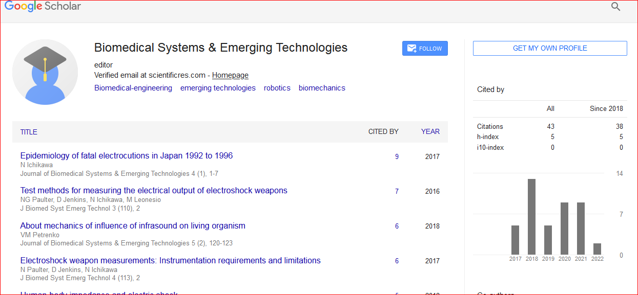
Citation: Peter, Ildiko. “Editorial Note on Biomedical Systems in Translational Orthopaedic Study.” J Biomed Syst Emerg Technol 9 (2022): 122. DOI: 37421/bset.2022.9.122
Copyright: © 2022 Peter I. This is an open-access article distributed under the terms of the Creative Commons Attribution License, which permits unrestricted use, distribution, and reproduction in any medium, provided the original author and source are credited.
Musculoskeletal disorder is a major burden on health care. Musculoskeletal complaints are the second most common reason for consulting a doctor, and constitute, in most countries, up to 10%-20% of primary care consultations. At any one time, 30% of American adults are affected by joint pain, swelling, or limitation of movement. According to the National Arthritis Data Workgroup, the best estimate of the national prevalence of arthritis specifically, osteoarthritis (OA), rheumatoid arthritis, low back pain, gout, and certain autoimmune connective tissue diseases was 15% in 1995. The total direct cost for use of health services that results from musculoskeletal conditions was 1.0% of the gross national product in Canada, and 1.2% in the USA. The indirect costs of musculoskeletal conditions (loss of productivity and wages) were much greater than the direct costs. Radiographic evidence of knee OA is prevalent in >30% of persons aged 60 years or older. It is expected that by 2020, this prevalence will increase to 20%, related in part to the advancing age of the population. Cartilage damage in OA is characterised as having an earlier dynamic phase, which is potentially reversible, followed by an irreversible pathological phase that ultimately leads to joint pain and immobility. The impetus to develop techniques to detect early lesions is to allow timely intervention to prevent the eventual evolution of radiographic joint space narrowing, osteophytosis, subchondral sclerosis, and cyst formation.
Animal models are an important part of orthopaedic research. They add to in vitro methods and provide the opportunity to study a specific biological mechanism in vivo. The ability to image a specific biological target in vivo can be translated into a part of orthopaedic diagnostic work-up. Commonly used techniques include Computed Tomography (CT), ultrasound, and Magnetic Resonance Imaging (MRI), single photon emission computed tomography (SPECT), and Positron Emission Tomography (PET). There is also an emerging imaging technology involving optical methods (fluorescence and bioluminescence) that are now used in preclinical animal models of disease. These particular tools are advancing the understanding and the related management of chronic musculoskeletal diseases, such as OA, rheumatoid arthritis, cancer, musculoskeletal pain, fracture healing, bone metabolism, chronic osteomyelitis, and osteoporosis. Through the use of multiple imaging modalities it is possible to study anatomy, physiology, and function in an in vivo model. Over the past several years, there has been significant development of dedicated instruments for small animal imaging applications in the modalities of MR, SPECT, PET, mCT, and in vivo optical imaging. MRI is a single imaging modality capable of high-resolution imaging, spectroscopy, and quantifying. Perfusion MRI using dynamic contrast enhanced MRI involves the rapid acquisition of serial MR images during and after administration of MR contrast agent. Based on the time following injection and the concentration of contrast in the tissues under investigation, a time intensity curve can be drawn allowing detection and quantification of wash-in and wash-out contrast kinetics. MR spectroscopy yields quantitative information on the chemicals that reside within the tissue.
The commonly measured elements in the musculoskeletal system are hydrogen, phosphorus, and sodium. Muscle diffusion-tensor imaging has been used for in vivo structural analysis and it has the potential to detect segmentation of muscle fibers in disease states. Blood Oxygen Leveldependent (BOLD) imaging was developed by Ogawa for functional MRI imaging for evaluating brain activation. This technique has recently been applied to evaluate oxygenation level in normal and diseased muscle. It has been reported that BOLD signal-based muscle functional MRI could be beneficial in understanding micro vascular-related disease such as muscular dystrophy, ischemia, and chronic/ peripheral venous insufficiency. SPECT and PET are functional imaging modalities that allow for molecular specific imaging in deep tissues of Nano and picomolar quantities, respectively. SPECT is a readily available technique for which there are a wide number of nuclear tracers available [1-5].
Google Scholar, Crossref, Indexed at
Google Scholar, Crossref, Indexed at
Google Scholar, Crossref, Indexed at