Opinion - (2022) Volume 8, Issue 4
Received: 01-Jun-2022, Manuscript No. JCTT-22-72348;
Editor assigned: 03-Jun-2022, Pre QC No. P-72348;
Reviewed: 09-Jun-2022, QC No. Q-72348;
Revised: 16-Jun-2022, Manuscript No. R-72348;
Published:
23-Jun-2022
, DOI: 10.37421/2471-9323.2022.8.182
Citation: Cartee, Todd V. “Hyperpigmentation on the Face and Crusty Papules.” J Cosmo Tricho 8 (2022): 182.
Copyright: © 2022 Cartee TV. This is an open-access article distributed under the terms of the Creative Commons Attribution License, which permits unrestricted use, distribution, and reproduction in any medium, provided the original author and source are credited.
Hyperpigmentation disorders are common dermatological conditions that can have substantial impact on quality of life. Recently, botulinum neurotoxin type A (BoNT-A) has been shown to be protective against UVB (type B ultraviolet rays)-induced hyperpigmentation in in vitro and animal models. This prospective, double-blind, randomized, controlled trial was carried out to investigate the effect of treatment with BoNT-A on subsequent UVB-induced hyperpigmentation in human subjects.
Fifteen healthy subjects older than 18 years old were enrolled in the study. Four separate areas of the abdomen were each randomized to receive intradermal injections of either 0.3 mL BoNT-A (IncobotulinumtoxinA; Merz Pharmaceuticals GmbH, Frankfurt am Main, Germany) at dilutions of 1:2.5, 1:5, 1:7.5, or normal saline (control). Fourteen days after the injections, experimental sites were irradiated by local broadband UVB to induce hyperpigmented spots [1]. The lightness index was measured at each experimental site using a colorimeter and hyperpigmentation scores were assessed using a 10-point visual analogue scale by a blinded physician and by study subjects [2].
Particulate matter with an aerodynamic equivalent diameter of 2.5 μm or less in ambient air (PM2.5) has become a global public and environmental problem and the control of the PM2.5 concentration in air is an urgent problem. PM2.5 can easily penetrate the skin, activating the inflammatory response in skin, unbalancing the skin barrier function and inducing skin aging. Hyperpigmentation is the main manifestation of skin aging and has a considerable impact on quality of life worldwide. To date, no research on the influence of PM2.5 on hyperpigmentation has been conducted. Here, we illustrate that PM2.5 can induce melanogenesis in vivo and in vitro by regulating TYR, TYRP1, TYRP2 and MITF expression via AhR/MAPK signaling activation. Furthermore, PM2.5 increased α-MSH paracrine levels, which in turn promote hyperpigmentation. Our results provide a deeper understanding of how PM2.5 disrupts skin homeostasis and function. Treatment with AhR antagonists may be a potential therapeutic strategy for hyperpigmentation induced by PM2.5.
The patient is a 57-year-old African-American woman who presented to the Vitiligo and Pigmentation Institute with a history of hyperpigmentation of the face, trunk and extremities. The skin discoloration had progressed during the 5 years preceding her evaluation at our institute. She had been treated for SLE with varying doses of HQ, from 200 to 400 mg daily and quinacrine for 16 years. At the time of evaluation, her SLE was in remission. Her medical history was unremarkable. Her initial SLE-related signs and symptoms included photosensitivity, alopecia, oral ulcers and arthralgias. She denied any history of bruising and had no risk factors for bruising, including any significance use of topical corticosteroids.
A 69-year-old woman with a history notable for lichen planus, breast cancer status post chemotherapy 7 years previously and fibroid-related menorrhagia status post hysterectomy and bilateral oophorectomy 20 years previously presented with a 6-year history of facial hyperpigmentation and new painful, nonpruritic, blisters, erosions and scabbing lesions on the backs of both of her hands. She had been taking ferrous sulfate 325 mg 3 times daily since her menorrhagia diagnosis 25 years previously. Exam revealed diffuse hyperpigmented patches on the face with hypertrichosis of the temporal region and fragile skin with crusted papules on the dorsal aspect of both hands [3-5].
Ashy dermatosis shares similar and distinct features with erythema dyschromicum perstans and lichen planus pigmentosa. These disorders were recently classified together within the spectrum of acquired macular pigmentation of uncertain etiology by a global consensus forum because of their significantly overlapping features. Although subtle variations in clinical presentation exist among these entities, they share histologic features of lichenoid changes, suggesting the role of the immune system in precipitating the classic hyperpigmentation. Consistent with these histopathologic features, ashy dermatosis has been associated with infections by parasites, enterovirus, hepatitis C and HIV. These viruses may result in infiltration of CD8+ T cells into the dermoepidermal junction, where they persist as a stable population. Subsequent exposure of CD8+ T cells to exogeneous stimuli such as medications or self-antigens may trigger an autoimmune reaction resulting in pigment incontinence that is characteristic of lichenoid diseases.
Intradermal BoNT-A was protective against subsequent UVB-induced hyperpigmentation in human subjects.
Crossref, Indexed at, Google Scholar
Crossref, Indexed at, Google Scholar
Crossref, Indexed at, Google Scholar
Crossref, Indexed at, Google Scholar
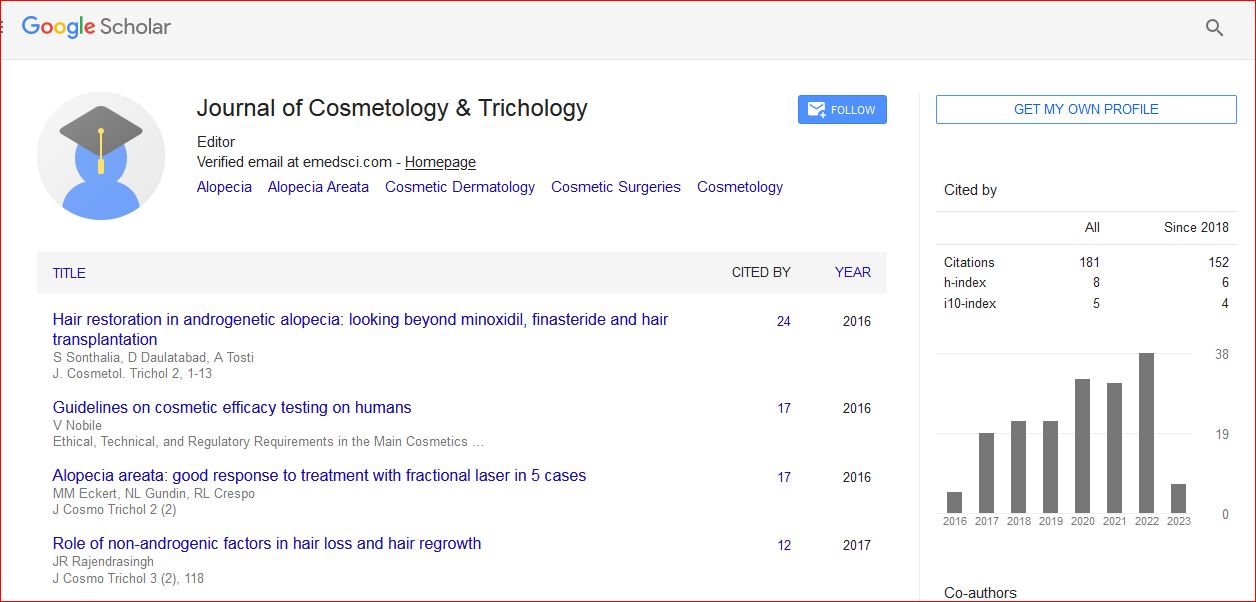
Journal of Cosmetology & Trichology received 180 citations as per Google Scholar report