Editorial - (2022) Volume 12, Issue 4
Received: 03-Apr-2022, Manuscript No. MCCR-22-67147;
Editor assigned: 05-Apr-2022, Pre QC No. P-67147;
Reviewed: 10-Apr-2022, QC No. Q-67147;
Revised: 15-Apr-2022, Manuscript No. R-67147;
Published:
20-Apr-2022
, DOI: 10.37421/2161-0444.2022.12.616
Citation: Dong, Kang. “Molecular Mechanisms of Action of Anticancer Drugs.” Med Chem 12 (2022): 616.
Copyright: © 2022 Dong K. This is an open-access article distributed under the terms of the Creative Commons Attribution License, which permits unrestricted use, distribution, and reproduction in any medium, provided the original author and source are credited.
All malignancies are caused by genetic abnormalities. Because of these mutations, critical proteins in particular signalling pathways are altered. Ras protein and p53 mutations are two of the most well-known and investigated modifications linked to malignant transformations. Almost all malignant changes are caused by mutations in Ras and p53 in the majority of instances. Approximately 20 to 30 percent of all human malignancies have mutated Ras genes. Ras proteins are switches that control cell proliferation, differentiation, and death, among other tasks. We now know that three types of genetic changes or mutations cause practically all malignancies. These mutations occur in oncogenes, tumour suppressor genes, and genes that control the precise replication of DNA, such as oncogenes, tumour suppressor genes, and oncogenes, tumour suppressor genes, and oncogenes, tumour suppressor genes, and oncogene Cellular checkpoint genes and DNA repair enzymes [1-5].
Histone Deacetylases (Hdacs)
Mutations are not necessarily the cause of changes in tumour suppressor genes or oncogenes. They could also be caused by epigenetic mechanisms such as DNA methylation and demethylation, as well as histone acetylation and deacetylation. The balance between histone acetylation and deacetylation, which is mediated by HATs and HDACs, is normally well regulated, but it is frequently disrupted in disorders like cancer. Conventional HDACs are made up of 11 members that require Zn2+ as a cofactor for deacetylase activity and are classified into four groups based on homology.
Histones are acetylated by HAT, which shifts their charge from positive to negative, reducing their interaction with negatively charged DNA. This makes the transcriptional apparatus more accessible, resulting in transcriptional activation. Deacetylation by HDACs can stop this chain of events from happening. Global transcriptional patterns can be affected by epigenetic alterations generated by imbalances between HATs and HDACs. Vorinostat is the most sophisticated HDAC inhibitor available for the treatment of advanced CTCL that has failed to respond to numerous or systemic therapies. Depsipeptide is a oneof- a-kind HDAC inhibitor prodrug that is transformed intracellularly to a reduced form with a functional sulfhydryl group capable of binding the zinc in class I HDACs' active site pocket.
Cisplatin
Cisplatin is a metallic (platinum) coordination complex having a square planar shape. It is also known as cis-diamminedichloroplatinum(II). At room temperature, it is a crystalline powder that is white, deep yellow, or yellow–orange in colour. It is soluble in dimethylprimanide and N,Ndimethylformamide, but very slightly in water. Cisplatin is particularly intriguing because it has been proven to have anticancer action in a range of tumours, including ovarian, testicular, and solid tumours of the head and neck. From a molecular standpoint, cisplatin is an excellent example of how a tiny change in Lung cancer is still one of the most common types of deadly malignancies. Platinum-based therapies are now the most effective therapy for SCLC. Two of the most frequent platinum-based therapies utilised in SCLC chemotherapy include cisplatin and carboplatin. Cisplatin is frequently used in clinical trials because of its potent anticancer properties, although it has side effects such as renal damage, nausea, and vomiting.
Urine volumes should be maintained to avoid renal damage, and large-dose infusion is required in cisplatin-based chemotherapy. Because severe hydration is often a concern, carboplatin has been considered a replacement for cisplatin in clinical practise with no apparent loss of therapeutic efficacy. The chemical structure of a target cell can have a big impact on its biological activity. In yeast and humans, the copper transporter Ctr1 is involved in cisplatin absorption. In vivo, knocking down CTR1 causes cellular resistance to cisplatin. Increased CTR1 expression leads to greater platinum buildup and, in most cases, increased cisplatin sensitivity. A membrane protein linked to cisplatin resistance in cells has been discovered.
Salicylic Acid
Salicylic acid has almost negligible inhibitory activity against pure COX, but it does suppress prostaglandin formation in intact cells, showing that it has anti-inflammatory benefits without inhibiting COX directly. The effects of aspirin and salicylic acid on COX-2 expression are controversial, with several studies suggesting a suppressive effect, no effect, or a potentiation of COX-2 expression. Aspirin and salicylic acid's activities on NF-B, which regulates the development of proinflammatory enzymes, cytokines, chemokines, immunoreceptors, and cell adhesion molecules, all of which play a role in inflammation and the immunological response, are the most commonly established COX independent activity. NF-B is generally stored in cells as inactive heterodimers complexed with inhibitor B (IB) proteins in a cytoplasmic protein termed inhibitor I-B kinase (IKK). IKK- and phosphorylate IB, splitting the IKK complexes into free NF-B dimers that translocate to the nucleus and activate genes involved in inflammation. At high doses, aspirin, salicylic acid, and sulfasalazine have been found to inhibit the function of IB kinase, preventing the release of NF-B and maintaining the IKK complexes.
Diallyl Disulphide
The organosulfur chemical diallyl disulfide (DADS) is generated from garlic and a few other Allium species. It is one of the main components of distilled garlic oil, along with diallyl trisulfide and diallyl tetrasulfide. It's a yellowish liquid with a strong garlic flavour that's insoluble in water. It is formed during the degradation of allicin, which is released when garlic and other Alliaceae plants are crushed. With hydrogen peroxide or peracetic acid, diallyl disulfide can easily be converted to allicin. There is evidence that DADS, by inducing phase-II detoxification enzymes, can protect humans from cancer. The tissue activities of QR and GT in the rat's gastrointestinal tract are increased by DADS. The glutathione (GSH) level of the intestinal mucosa and liver can be effectively increased by DAD.
Glutathione S-transferase A5 (rGSTA5) and aflatoxin B1 aldehyde reductase 1 are both induced by DADS (rAFAR1). The key mechanism by which DADS protects against aflatoxin B1 (AFB(1))-induced carcinogenesis is the induction of rGSTA5 and rAFAR1. Through adjustment of their internal redox environment, DADS may decrease the proliferation and arrest of the G2/M phase in human colon cancer HT-29 cells. Upstream of p53 activation, ROS trigger the DADSinduced cell-cycle arrest implicated in stress-induced signalling. In human colon cancer cells, DADS reduces the percentages of viable cells in a dose- and time-dependent way, causing the G2/M phase to be arrested, which is connected with the formation of ROS and an increase in the expression of cyclin B.
None.
The author reported no potential conflict of interest.
Google Scholar, Crossref, Indexed at
Google Scholar, Crossref, Indexed at
Google Scholar, Crossref, Indexed at
Google Scholar, Crossref, Indexed at
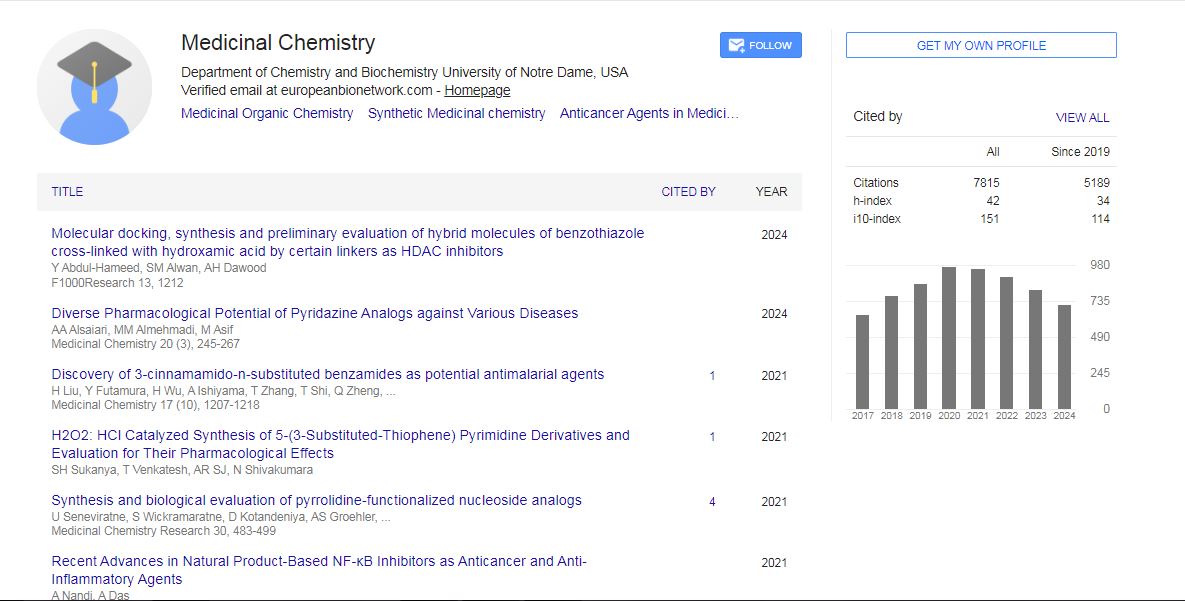
Medicinal Chemistry received 6627 citations as per Google Scholar report