Commentary - (2022) Volume 12, Issue 2
Received: 03-Mar-2022, Manuscript No. JBL-22-60493;
Editor assigned: 08-Mar-2022, Pre QC No. JBL-22-60493;
Reviewed: 19-Mar-2022, QC No. JBL-22-60493;
Revised: 26-Mar-2022, Manuscript No. JBL-22-60493;
Published:
02-Apr-2022
, DOI: 10.37421/ 2165-7831.2022.12.283
Citation: Romanoff, Selma. "Note on Diagnoses of Left Ventricular Thrombus ". J Blood Lymph 12 (2022): 283.
Copyright: © 2022 Romanoff S. This is an open-access article distributed under the terms of the creative commons attribution license which permits unrestricted use, distribution and reproduction in any medium, provided the original author and source are credited.
Left Ventricular Dysfunction is closely linked to LVT. In a study of patients with LVT, all had some form of Left Ventricular Disease: ischemic cardiomyopathy in patients, dilated cardiomyopathy, stress-induced cardiomyopathy, apical hypertrophic cardiomyopathy, severe aortic stenosis, and cardiac arrest in one patient each. Recent Myocardial Infarction is the most common risk factor for LVT. LVT was seen in of all patients after an acute MI in the pre-thrombolytic era with anterior MI. Primary percutaneous coronary intervention is linked in LVT post-acute MI and with anterior MI. LVT, on the other hand, can develop in people who have no known heart illness. LVT was found in patients with cerebral ischemic stroke or Transient Ischemic Attack (TIA) with sinus rhythm without a history of cardiac illness.
LVT is a dangerous but uncommon consequence of acute MI that is linked to systemic thromboembolism, which can lead to embolic strokes. The majority of patients diagnosed with an acute MI should have their left ventricular function evaluated, including the apical function of the left ventricle and the presence of an LVT. The conventional transthoracic echocardiogram is the most commonly utilized diagnostic modality. When the left ventricular apex is difficult to see on a TTE due to the patient's body habitus, and there are anterior or apical wall-motion abnormalities with a high apical wall-motion score (>5 on noncontrast TTE), contrast TTE or cardiac MRI should be considered, depending on local availability and resources.
OAC therapy should be begun right away in patients who have been diagnosed with an LVT. Guidelines are still young and regularly changing due to a lack of randomized control evidence in this field. OAC may be explored in patients with STEMI who have anterior apical akinesis or dyskinesis, according to the 2013 American College of Cardiology Foundation/American Heart Association STEMI Guidelines, to prevent LV thrombus formation. Anticoagulation may be considered for 3 months in patients with acute anterior STEMI and ischemic stroke or transient ischemic attack who have anterior apical akinesis or dyskinesis, according to the AHA/American Stroke Association 2014 Guidelines on Stroke Prevention.
Once an LV thrombus is diagnosed, the European Society of Cardiology's 2017 STEMI Guidelines indicate that OAC be explored for up to 6 months, guided by recurrent echocardiography and taking into account the risk of bleeding and the necessity for concurrent antiplatelet medication. As a result, the ideal duration of OAC in these individuals is unknown, and choices about whether or not to continue OAC should be determined on a case-by-case basis.
Patients with signs of LVT after MI are frequently prescribed warfarin in addition to DAPT. Patients should be bridged with a parenteral anticoagulant until a therapeutic INR (2.0-3.0) is obtained for at least 24 hours, due to the significant risk of thrombosis and stroke. While warfarin is the most regularly used OAC and the agent with the longest history of use, it has a number of drawbacks, including the need for frequent monitoring, a narrow therapeutic window, dietary restrictions, and various drug-drug interactions.
Because of these issues, the use of direct oral anticoagulants for this condition is becoming more common. Finally, in 2014, the American Heart Association/American Stroke Association Guidelines on Stroke Prevention recommended that lowmolecular- weight heparin, dabigatran, rivaroxaban, or apixaban be considered as an alternative to vitamin K antagonists for post- MI LV thrombus or anterior or apical wall-motion abnormalities with an LV ejection fraction less than 40% who are intolerant of vitamin K antagonists due to nonhemorrhagic.
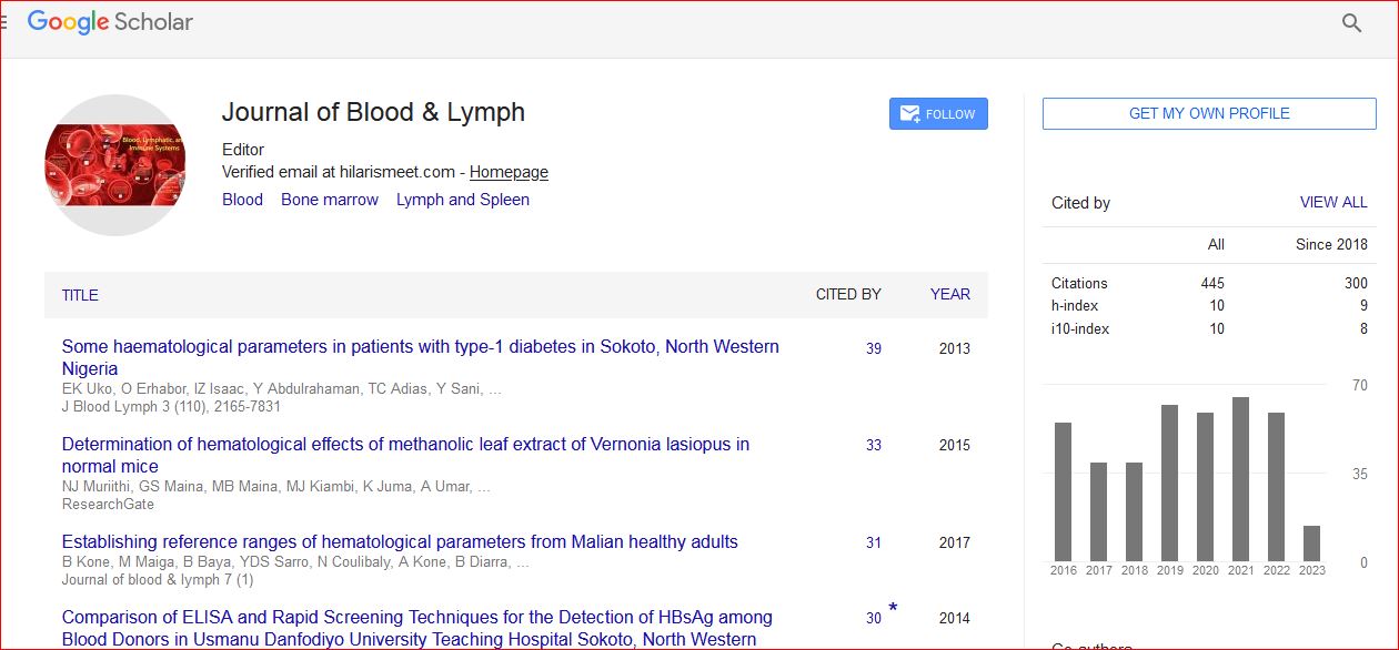
Journal of Blood & Lymph received 443 citations as per Google Scholar report