Research Article - (2022) Volume 6, Issue 3
Received: 16-Apr-2022, Manuscript No. ahbs-22-61028;
Editor assigned: 18-Apr-2022, Pre QC No. P-61028;
Reviewed: 02-May-2022, QC No. Q-61028;
Revised: 07-May-2022, Manuscript No. R-61028;
Published:
14-May-2022
, DOI: 10.37421/ahbs.2022.6.161
Citation: Itefa, Tesfaye, Yoseph Alemu, Ebisa Regasa and Amanuel Alemu, et al. “Prevalence of Fasciolosis (Liver Flukes) Infection in Cattle Slaughtered at Dambi Dollo Town Municipal Abattoir, Kellem Wollega Zone, Ethiopia.” J Anim Health Behav 6 (2022): 161
Copyright: © 2022 Itefa T, et al. This is an open-access article distributed under the terms of the Creative Commons Attribution License, which permits unrestricted use, distribution, and reproduction in any medium, provided the original author and source are credited.
The study was carried out from January to December 2021 with the main objectives of to determine the prevalence of fasciolosis infections in cattle, slaughtered at Dambi Dollo Municipal Abattoir. The slaughtered animals were daily inspected for liver fasciolosis throughout the year of 2021. Macroscopic fasciolosis was detected from a total of 4424 basing on animals species, sex, season, and Fasciola species. In addition to this, fecal samples from 100 female cattle were collected for microscopic examination. The total prevalence rate of Fasciola species infection occurs in the study area were about 1364/4424 (30.83%) from the total cattle slaughtered carcasses. On sex based case, prevalence of fascioliasis was 8/24 (33.33%) and 1356/4400 (30.82%) for females and males cattle carcasses, respectively. The study revealed that the significance of season in finding that highest fasciolosis infection was recorded during winter and autumn. It constitutes a major cause of economic losses at study area and threat public health.
Fasciola gigantica • Fasciola hepatica • Liver fluke • Slaughterhouse • Snails
Slaughterhouses provide an excellent meat inspection place, where many zoonotic diseases observed but meat poor handling in or out the abattoir can leading to both economic losses and a lot of public health hazardous [1,2]. Fasciolosis considered the top of all the domestic ruminants’ parasitic zoonotic worldwide infection that is endemic in a tropical area and Kellem Wollega, Dambi Dollo [3-5].
Genus Fasciola “liver fluke” is belonging to trematode helminths which containing two main species; Fasciola gigantica and Fasciola hepatica is very common observed in the liver of cattle and other ruminants [6-8]. Fasciolosis reduces animal productivity, weight gain, and the production of meat and milk. In addition, it causes moderate icterus, metabolic disorders, and secondary infections due to decrease immunity by chronic fasciolosis and liver condemnation during postmortem inspection in slaughterhouses while the acute fasciolosis may lead to mortalities [9-11]. Human fasciolosis infection occurs accidentally after ingestion of eggs/larvae while ruminant ingestion of forage containing metacercarial cyst [12].
Ingested parasite lives in hepatic parenchyma or in bile duct, which causing liver hemorrhagic black tunnels [13]. Diagnosis is depending on the history of snail habitats or fasciolosis on the farm, symptoms, postmortem examinations, feces, and blood examination for Fasciola eggs [14].
There is no enough information on the ruminants’ fasciolosis in the study area, Dambi Dollo town. Therefore, this study was designed with the aims of determining the prevalence of fasciolosis infections in cattle slaughtered in Dambi Dollo town slaughterhouse.
The study area
A cross-sectional study was conducted in Dambi Dollo Municipal abattoir to detect the prevalence of the fasciolosis (liver flukes) from the slaughtered cattle. Dambi Dollo town is the capital city of Kellem Wollega Zone Administration. It is a part of the Oromia region, which is located to the west of the Addis Ababa/Finfinne between 8°32°N latitude and 34°48°E longitude with an elevation between 1701 and 1827 meters abov sea level. Dambi Dollo town is located 652 km to the west of Addis Ababa/Finfinne. Dambi Dollo Municipal abattoir slaughtered about 4424 cattle animals during 2021. According to the Ethiopian legislations of meat inspection, slaughtering of female cattle never been allowed before all teeth are changed (over 5 years) while they approved for slaughtering after about 2 years.
Samples collection
A total of 4424 (4400 bulls and 24 cows) local breed cattle slaughtered at Dambi Dollo Municipal abattoir were inspected for the presence of liver fluke/fasciolosis allover 2021 which was efficiently inspected by naked eye and palpation for the presence of gross lesion and the worms, then further examinations done at Laboratory level. All data samples collected were transported in an icebox to the laboratory of Type B Veterinary Clinic of the town for further examinations within 24 hrs.
Samples preparation for postmortem inspection
Liver and gall bladder postmortem inspection by making multiple cuts and sub cuts about 1 cm thick to check the presence of fasciolosis, which made gritty sounds and bile duct thickness, palpation pressure, exerted brownish fluid, and immature Fasciola. Identification of the species based on the morphological features of the agent and classify into F. gigantica and F. hepatica [15,16].
To calculate the total sample size, the following assumptions were made: 5% desired level of precision, 95% level of confidence, and 50% expected the prevalence of cattle fasciolosis in Dambi Dollo Municipal abattoir, the sample size was determined by using the formula given below [17].
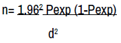
Where,
n = required sample size,
Pexp = Expected prevalence,
d=desired absolute precision. But in current stud, all slaughtered animal samples were taken for good precision.
Statistical analysis
The obtained results were encoded and recorded in an excel database analyzed by descriptive statistics survey were performed using Graph Pad Instant version 3 for determination of means and the analysis of variance between the different data. The treatment, in this study, was determined using standard error and analysis of variance (p<0.05). The incidence of fascioliasis was 8/24 (33.33%) and 1356/4400 (30.82%) for females and males cattle carcasses, respectively.
Seasonal liver fascioliasis condemnation rates in examined cattle and buffaloes samples
As illustrated in Table 1, results revealed that cattle’s fascioliasis is higher during spring and summer. The highest fasciolosis infection found in winter followed by autumn, spring, and summer. There was a significant difference in between different seasons while there was not any significance between males and females cattle. The highest fasciolosis infection found in winter followed by autumn, spring, and summer. There was a significant difference in between different seasons. The cattle fasiolosis prevalence rate was (35.04%, 22.73%, 18.48%, and 23.75%) during winter, spring, summer, and autumn, respectively.
| Season | Winter | Spring | Summer | Autumn |
|---|---|---|---|---|
| Rate of infection | 35.04 | 22.73 | 18.48 | 23.75 |
Macroscopic liver fasciolosis in examined cattle samples
Grossly regarding fasciolosis infection during slaughterhouse postmortem inspection (Table 1) showing the external smooth liver surface declared several white or creamy tunnels ranged from few millimeters to nearly 3 cm, represented the postmortem liver fibrosis appear from external liver surface. Fasciolosis tunnels which observed from intact liver surfaces oozing grassy blackish hemorrhagic exudates. Creamy leaf-like Fasciola spp. about 1.5-2.0 cm in length and about 1.0 cm in width were observed by naked eye from the liver of the slaughtered cattle (Table 2).
| Examined Animals | Examined | Positive | Prevalence% | Overall Prevalence% |
|---|---|---|---|---|
| Cattle | - | - | - | - |
| Females | 24 | 8 | 33.33 | - |
| Males | 4400 | 1356 | 30.82 | - |
| Total | 4424 | 1364 | - | 30.83 |
Prevalence of liver fasciolosis in examined cattle samples
The results obtained in Table 2 indicated that the overall prevalence rate of Fasciola infection occurs in the study area were 1364/4424 (30.83%) from the total slaughtered cattle carcass.
The sex based prevalence of fasciolosis was 8/24 (33.33%) and 1356/4400 (30.82%) for females and males cattle carcasses, respectively (Table 1).
Fasciola spp. is a parasite threatening domestic ruminants and public health. Transmission of this trematode infection is depending on the presence of intermediate “lymnaea snail” host and final host. This snail host commonly presents in high density during rainfall period annually and/or in highly moist pastures soil [13,18]. The overall prevalence rate of fasciolosis in the examined cattle slaughtered in Dambi Dollo town municipal abattoir was about 1364/4424 (30.83%) which nearly agreed with Morsy TA, et al. [19], who previously found 25.5% in Egypt. On the other hand, higher incidences of fasciolosis have been recorded by Pfukenyi DM and Mukaratirwa S [20], who reported 37.1% in Zimbabwe and Abraham JT and Jude IB [13] recorded 44.8% in Nigeria. However, there were some remarkable lower results reported by Mellau LSB, et al. [21], who found 16.3% in Tanzania, Haridy FM, et al. [22] noted 21.8% in Gambia Governorate, Afrakhosravi EB [23] reported 11.09% in Iran, and Mungube EO, et al. [24] recorded 26% in Kenya. Human fasciolosis was been occurred after the consumption of encysted cercaria and not by eating of animal livers infected by adult Fasciola spp.
The ingestion of watercress vegetables grown along contaminated water by snails and domestic ruminant fecal matters with adult parasite [25]. Our reported seasonal liver fascioliasis condemnation rates revealed that is lower during winter and autumn than in spring and summer.
Accordingly, the study found that (35.04%, 23.75%, 22.73%, and 18.48%) during winter, autumn, spring, and summer, respectively. This finding might be attributed to raining season and presence of fresh green grazing pasturing. This finding was supported by the previous findings reported by Adedokun OA, et al. [26] who reported in winter (52.3%) and in dry season (21%) in Nigerian cattle, while, fasciolosis was highest in winter (around the raining periods) and/ or dampness area due to spreading of the snails host [13,23,27,28].
Fasciolosis occurs mainly not only in children living in rural settings but also in people living in urban areas by metacercarial of the fluke is ingested along with watercress salad and vegetables grown along banks of water reservoirs inhabited by potential snail hosts. About 2.4 million people infected world wide and 180 million are at risk of the infection fasciolosis commonly asymptomatic children infection with mild anemia. Humans’ fasciolosis is mainly correlated with highly eggs excreted areas and not related with highly animals’ fasciolosis and sometimes infection transmitted by human stool contamination [29].
In this study, the routine macroscopic postmortem fasciolosis inspection revealed that infected liver have numerous injuries with congestion, enlargement with very hard fibrosis. Postmortem visually examination of intact liver also showing the presence of different sizes (1.5-2.7 cm) of Fasciola spp. impeded on the hepatic tissue with characteristic white or creamy color. Hepatic postmortem incision is showing thick wall fibrosis by fasciolosis tunnels which oozing grassy blackish exudates and debris. The trials to opening this tunnel exerted leaf-like liver flukes that diminished infected liver and carcass value and resulted in rejection of liver by consumers.
Similar lesions were observed by authors in Bangladesh [18] and in Nigeria [2,13]. According to Ethiopian veterinary authorities, detection of fasciolosis in liver should be removed total liver condemnation or partial affected lobes after performing boiling tests and rapid phase according to parasitic infestation density and extension. The rest carcass was been released for human consumption [25]. Controlling fasciolosis mainly by anthelmintics, this only acts against at mature stages. Triclabendazole is the only drug, which affects against both immature and mature stages fascioliasis. Anthelmintic administered during December/January and from April/May for controlling chronic fasciolosis, a third dose should be given in August. However, molluscicides were been recommended for snail control [20,30].
The present study revealed a moderate fasciolosis infestation in cattle in the municipal abattoir of Dambi Dollo town, and the study is recommended that it is important to enhance snail and fasciolosis control at farm levels to diminish the economic losses due to infection. Thorough meat inspection should also be taken on abattoir by experts.
Google Scholar, Crossref, Indexed at
Google Scholar, Crossref, Indexed at
Google Scholar, Crossref, Indexed at
Google Scholar, Crossref, Indexed at
Google Scholar, Crossref, Indexed at
Google Scholar, Crossref, Indexed at
Google Scholar, Crossref, Indexed at
Google Scholar, Crossref, Indexed at
Google Scholar, Crossref, Indexed at
Google Scholar, Crossref, Indexed at
Google Scholar, Crossref, Indexed at
Google Scholar, Crossref, Indexed at
Journal of Animal Health and Behavioural Science received 38 citations as per Google Scholar report