Perspective Article - (2022) Volume 13, Issue 5
Received: 02-Sep-2022, Manuscript No. hgec-23-95631;
Editor assigned: 05-Sep-2022, Pre QC No. P-95631;
Reviewed: 17-Sep-2022, QC No. Q-95631;
Revised: 22-Sep-2022, Manuscript No. R-95631;
Published:
29-Sep-2022
, DOI: 10.37421/2161-0436.2022.13.189
Citation: Otterloo, Eric. “The Base of the Skull: An Overview of the Cranial Base in Brief.” Human Genet Embryol 13 (2022): 189.
Copyright: © 2022 Otterloo E. This is an open-access article distributed under the terms of the Creative Commons Attribution License, which permits unrestricted use, distribution, and reproduction in any medium, provided the original author and source are credited.
The core of the cranium, from the rostral to caudal ends, is the multipurpose bony platform known as the cranial base. It serves as a link between the head and the vertebral column below, supports the brain and skull vault, and seamlessly integrates with the facial skeleton at its rostral end. The cranial base, which is distinct from the majority of the cranial skeleton, is formed by endochondral ossification from a cartilage intermediate known as the chondrocranium. Anomalies of the cranial base frequently coexist with congenital birth defects affecting these structures due to the close connection of the cranial base to nearly all aspects of the head. In spite of this crucial significance, research into the genetic control of cranial base development and the disorders that are associated with it lags behind that of other craniofacial structures. Here, we feature and audit formative and hereditary parts of the cranial base, including its change from ligament to bone, double embryological beginnings, and vignettes of record factors controlling its development.
Despite being concealed within the cranial core, the cranial base almost functions as an anatomical girder. The viscerocranium, or facial skeleton, largely from its rostral end, while the cranial base houses the brain and the cranial vault that lies above it. The cranial base develops almost to accommodate, protect, and support these specialized organs because of its proximity to and co-development with the optic and nasal placodes, which are cranial sense organs. Additionally, the cranial base is traversed by numerous nerves and blood vessels through its numerous foramina due to its anatomical location within the centrum of the head, just inferior to the brain. The completed cranial base, like the cranial vault above it, is almost entirely made up of tissues that come from mesodermal or ectomesenchymal neural crest cells. Notwithstanding, as a glaring difference to the calvarias or the brain peak cell determined viscerocranium which go through a course of intramembranous solidification the cranial base carries out a 'long bone methodology' including endochondral hardening. The cranial base's embryonic development begins as a collection of distinct cartilages that combine to form a single that eventually undergoes endochondral ossification. Especially at a high level of molecular resolution, our comprehension of the cranial base in relation to the easily accessible viscerocranial or neurocranial calvarial elements is lacking This is in part due to the central position of the cranial base within the skull. However, developmental anomalies affecting the cranial base frequently manifest viscera- and neurocranial defects due to the cranial base's proximity to visceroand neurocranial elements. As a result, gaining a better understanding of this occluded structural component will provide a comprehensive understanding of the formation, disruption, and evolution of the craniofacial complex as a whole [1].
Methanol and ethanol blends with diesel fuel have a density. This enables their delivery to the diesel cylinder using fuel pumps and injector nozzles intended for the primary fuel Methanol and ethanol are both extremely hazardous flammable liquids in their purest form, with a flash point and an ignition temperature of respectively. Methanol has a lower caloric value but its gains over diesel fuel are offset by its higher combustion rate, full combustion, and lower heat losses. Pure methanol and ethanol, however, are exclusively utilised as fuel in diesel automobile engines. Blends of petroleum fuel, methanol, and ethanol must undergo continuous hydrodynamic treatment [2].
Artificial liquid fuel with biological origins is one of the popular and promising types of alternative fuel known as methyl esters of fatty biodiesel fuel is produced from a variety of sources, most notably rapeseed, plants, and used cooking oil. The availability of vast quantities of raw materials is a benefit of using biodiesel fuel. Diesel and biodiesel can be combined or used separately. Additionally, it should be noted that the use of alternative fuels is restricted for high-power marine diesel engines cylinder diameters greater than rated powers greater than because doing so results in uncontrollable environmental performance degradation and a reduction in effective power at the rated load [3].
The cartilage anlage of the cranial base, which is a subset of the chondrocranium and has been extensively, discussed elsewhere consists of cartilage elements and grows close to the viscerocranium and These ligament components follow still up in the air and -transiently arranged formative program, including their amazed appearance from undifferentiated mesenchyme into recognizable, dense, structures. The elements of the caudal region, such as parachordal cartilages, first appear. Second, the rostral elements become apparent. Finally, a continuous "cartilaginous platform" runs from rostral to caudal during midline Additionally, cartilaginous such as the ala temporalis cartilage, and lateral elements, such as the parietal and frontal cartilages, emerge from or near this core structure, eventually joining the inferior lateral components of the cranial vault. Another important function of the cranial base is to encase, protect, and support the nasal and auditory apparatus cranial sense organs. The mouse's chondrocranial development process begins close to embryonic day all of the cartilage's individual components have fused into a single structure. Again, unlike the other intramembranous components of the craniofacial skeleton, which do not utilize a cartilage intermediate, this intermediate cartilage template is formed in a unique manner [4,5].
The physician faces a number of obstacles when treating couples. To explain the scope of the diagnostics performed, which ultimately lead to targeted therapies, a standardized and structured diagnostic procedure is required. However, these clear strategies are obscured by various guidelines' recommendations. After all, different guidelines have very different times to start diagnostic procedures. After two clinical pregnancies in a row, it seems reasonable to conduct diagnostics of the most common causes, as stated in recommendation. While guidelines speak of three consecutive pregnancy losses, the guidelines also include two non-consecutive losses. Therefore, it is essential to establish a workflow that takes into account additional circumstances. A recent meta- was unable to demonstrate a difference in the prevalence of uterine anomalies or an among women who had lost two or more pregnancies.
None.
None.
Google Scholar, Crossref, Indexed at
Google Scholar, Crossref, Indexed at
Google Scholar, Crossref, Indexed at
Google Scholar, Crossref, Indexed at
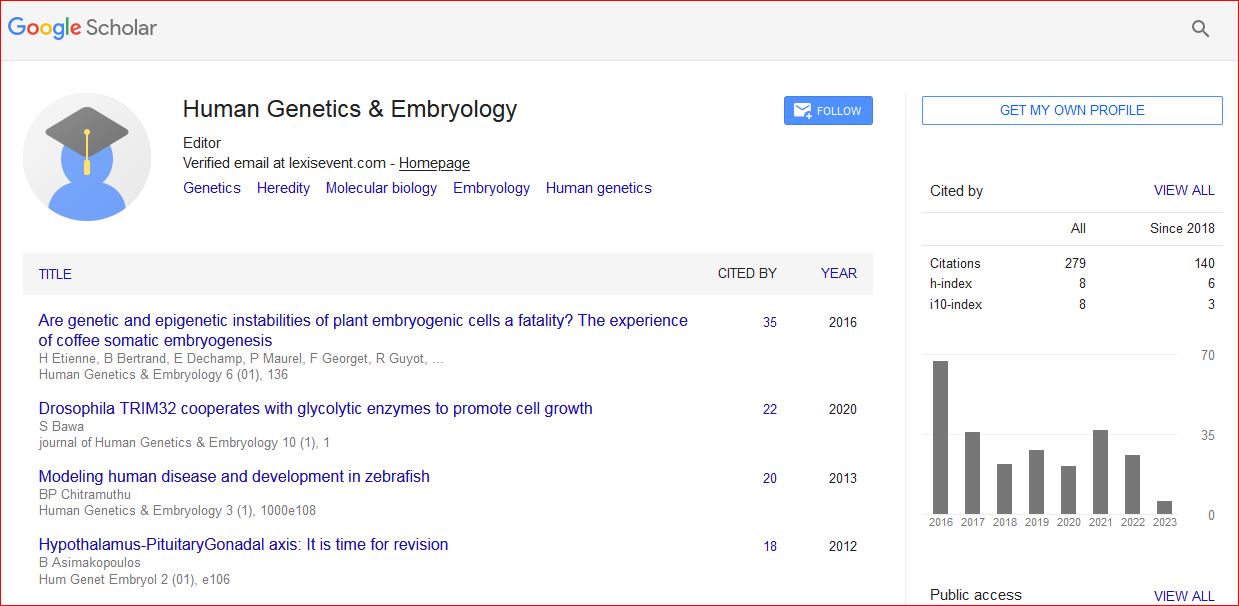
Human Genetics & Embryology received 309 citations as per Google Scholar report