Research Article - (2024) Volume 15, Issue 2
Received: 01-Apr-2024, Manuscript No. jvst-24-132078;
Editor assigned: 03-Apr-2024, Pre QC No. P-132078;
Reviewed: 16-Apr-2024, QC No. Q-132078;
Revised: 22-Apr-2024, Manuscript No. R-132078;
Published:
30-Apr-2024
, DOI: 10.37421/2157-7579.2024.15.236
Citation: Mohammed, Asledin. “The Study on Occurrence
of Mange Mite Infestation on Camel, Zoonotic Important, Economic Losses with
Associated Risk Factors in Pastoral Areas of Kumbi District East Hararghe Zone,
Ethiopia.” J Vet Sci Technol 15 (2024): 236.
Copyright: © 2024 Mohammed A. This is an open-access article distributed under the terms of the Creative Commons Attribution License, which permits unrestricted use, distribution, and reproduction in any medium, provided the original author and source are credited.
A cross-sectional study was conducted from July up to december 2024 to in Kumbi district, Eastern Hararghe zone, Oromiya regional state. The objective was by implementing study design to determine the prevalence of mange agents involved in camel skin diseases and their host risk factors. Mange mite most important parasitic diseases of livestock by affecting skin and hide as well as decrease quality and quantity of production. Camel mange is economically important contagious camel disease that has impact on their productivity and health. For clinical examination 384 camels was randomly selected and examined by implementing skin scrapings Procedures under microscope. The prevalence of infected camel from examine Was 25%. Only Sarcoptes was identified as the only mite species in all skin scraping samples collected from the suspected mange mite lesions as well as zoonotic problem. The variation in prevalence of mange mites (P<0.05) is depending on origin, sex, age and body condition. Inorder to avoid economic by impact of mange on camel production, consequently, strategic mange control with specific management should be implemented.
Prevalence • Management • Risk factor • Camel • Kumbi • Skin scraping
Camels play an important socio-economic role in the arid and semi-arid areas, where most of the resource poor farmers in Africa live. World camel population is estimated to be around 25.89 million across 47 countries. About 85% of the camel population inhabits mainly eastern and northern Africa and the rest in Indian subcontinent and Middle East countries [1]. A well-known feature of camel is its ability to survive and produce in drought areas where other animals hardly survive and that is the reason why there is a steady increase in the number of camel while the recurring drought is causing huge losses in other livestock species in the horn of Africa [2]. Ethiopia is one of the largest camel populated countries in the world. In Africa, it ranks third next to Somalia and Sudan. Its ability to withstand torrid heat and extreme desiccation are of paramount importance in determining its distribution. The normal distribution of the camel is in the Africa and Asian subtropical dry areas. The Eastern and southern parts of the country namely, Afar, Somali and Borena are the major areas were camel husbandry is widely practiced. In this area, the livelihood of pastoral communities is certainly ensured by dromedaries. Camels are an important source of milk, meat and their dung is used for fires. They are also used for transport purpose; furthermore, camels are exported mainly to Egypt and Sudan, and are also slaughtered for meat consumption during ritual occasions [3]. Despite the fact that, camels provide lots of socio-economic advantages and are the preferred domestic animal species in the ever-changing climate, so far it was neglected by researchers and development planners [4]. Several endo and ectoparasites have been identified as the major problems affecting the health, productivity and performance of camels [4,5]. Although camels were considered in the past, and for a fairly long time, as resistant to many disease causing factors, it has been proved that camels are susceptible, the same as other livestock or even more, to the common disease causing pathogens affecting other animal species [6]. Pathogenic diseases, poor nutrition and traditional management systems as well as lack of veterinary services have hampered their full utilization, despite the importance of dromedary in the semi-arid and arid areas where the environment is harsh and hostile [7].
In Ethiopia Camel mange, an extremely contagious ectoparasitism caused by the parasitic mite Sarcoptesscabiei and transmitted by direct or indirect contact, is one of the most important parasitic diseases affecting camel. Slow reproduction cycle, high calf mortality and health problems are major constraints in increasing camel herd population and productivity. Ectoparasites (mites, ticks and insects) of the camel and their capacity to disease transmission are important constraints to productivity and performance [2]. The economic values of mange infested animal emanate from decreased body weight, expense of therapy, deterioration of skin due to perforation of the skin and intense pruritus as skin lesions may cover almost the entire body, and occasional mortalities in untreated and young animals. In addition, mange mite has enormous zoonotic and public health significance.
Knowledge about the prevalence of the diseases together with associated risk factors as part of the epidemiology of the disease is crucial for any attempt of prevention and control of the disease in question. Even though, there were several works done in other species of animals, there was no any study on the prevalence and risk factors associated with camel mange in Kumbi Woreda. Therefore, the objectives of this study were:
Economic and zoonotic importance of camel mange
The economic values of mange infested animal emanate from decreased body weight, expense of therapy, deterioration of skin due to perforation of the skin and intense pruritus as skin lesions may cover almost the entire body, and occasional mortalities in untreated and young animals. Moreover, mange can harshly decrease the welfare of milking animals as reducing the vitality and increasing susceptibility to other diseases as a result of secondary bacterial infection. It can abridge milk production and disserve milking procedure as a result of uneasiness of infested animals. During development of mange, itchiness distracts the animals from eating so that they often become emaciated. The specific lesions are confined to the integument and comprise hyperkeratosis, anaemia, general loss of productivity and body weight.
The scabies or skin scabs are a major public health problem of poverty hit regions in the world since 1687. The disease is caused by a small mite Sarcoptesscabiei which burrows into the epidermis giving it a look of the short wavy line. The lesions include the development of papules which later on develop into vesicles, excoriations, eczema, secondary infections and crusts and the symptoms are commonly visible in inter digital spaces of the hands, wrists, penis, face and neck. The animal scabies is self-limiting in humans as the mites cannot complete their life cycle. The disease spreads to humans, especially the animal handlers through direct contact with the diseased animals or becoming in contact with fomites of animals and produces pruritic papules and itch in humans. The transfer of disease, from a camel to man may usually take place during milking, handling or riding. Treatment of both animals and the camel handlers can help in controlling this zoonotic problem.
Description of study area
The study was conducted in selected peasant associations in Hararge district of Kumbi woreda, Eastern Ethiopia. The area was located 814 kilometers east of Addis Ababa and 308 km away from the city of Harar. It is sarowunded by Gola Oda, Mayu Muluke, Burka dhimtu. The annual Minimum and Maximum temperatures are 16 and 28°C, respectively. Agro-climatic condition of the area is semi-arid and arid with mean Annual rain fall 1300 mm. The rainy seasons in the area from June to September wich was used for crop production, pasture and water harvest and the short rains season from February to May mainly used for land preparation, planting of long cycle crops collected after the june to September several rains, small scale production, and improving water and pastures. Total livestock population of Kumbi districts are 696,440; of which 206678 Cattle, 191444 goats, 140238 sheep, 19152 donkeys, 43 mules, 131857camels and 4488 poultry and 2540 Bee hives.
Study design and period
A cross sectional study was used to estimate the prevalence and associated risk factors for the occurrence of camel mange in the study area between April and November 2024. The study animals were sampled by simple random sampling from selected Peasant associations located in Kumbi Woreda based on the accessibility study population, willingness of the camel holder, considering their settlements, road accessibility and transport.
Study population and sample size determination
The study animals were indigenous breeds of one humped camel (Camelus dromedaries) reared under pastoral management system in free grazing, and usually mixed with livestock from other districts, the animals move from feed shortage area to feed abundant areas especially during drought season. All age and both sex of camels were included categories in this study.
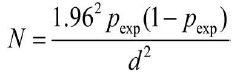
Collection Skin scrapings of 384 camels with considering of peasant association, age, sex, body condition, and herd size, was taken from different camel populations in Kumbi woreda selected 3 kebele. Age of studied camels was categorized into <3, as a young and >4 years as an adult camel. The age of the sampled animals was determined by dental eruption according to FAO [1]. The Body Condition Score (BCS) of sampled camels was evaluated by looking the back and flank then categorized as good, medium and poor according to Faye B, et al. [8]. Herd sizes as small (less than twenty), as medium (between twenty and forty) and large (greater than forty) were determined according to classification of Megersa B, et al. [9]. After selection of animals, each camel was restrained properly and the hairs were shaved using scalpel blade from the edges of the lesions till blood oozes out of the capillary.
Examination of mange mites investigation
Skin scrapings from suspected cases of mange were collected in labelled Petri-dishes and preserved in 10% formalin and taken to laboratory and 10% potassium hydroxide (KOH) was added to digest or clean the scraped material of skin, hair, and other debris so that mites released from scabs and crusts before examination following procedures indicated by Soulsby. All scraped tissues were carefully placed on microscopic slide for microscopic examination (10 x or 40 x magnifications) and identification of the mange mite species based on the morphological characteristics described by Urquhart.
Clinical examination and scoring of skin lesions
All camels were subjected to whole body examination for clinical signs of mange (erythema, pruritus, alopecia, hyper pigmentation and crusting) and clinically scored according to a system previously applied to horses with chorioptic mange [10]. Prior to enrolment in the study, the camels were tested for presence of mites (larvae, nymphs and adults) in skin scrapings obtained from at least 3–4 sites, the severity of skin lesions mild and moderate with recovery.
Statistical analysis
Microsoft excel spread sheet program was used to store all the data and Statistical Package for Social Sciences (SPSS) version 22.00 software was used to analyse the data. Prevalence of mange mites was computed as the number of each sample items positive for mange divided by total number of the samples examined. Chi-square (χ2) was used to test the presence of association between variables. When P value was less than 0.05, the presence of significance difference was considered.
The overall prevalence of camel mange mites in this study was found to be 25% (96/384). In this study, only sarcoptes scabiei var. cameli was identified as the only mite species in all skin scraping samples collected from the suspected lesions. Prevalence of Camel Mange based Peasant Associations In the study area, Urgo and Ija-Godda were found with slightly higher prevalence followed by Kara-Balchi having 28.1%, 24.6% and 22.1% respectively. There is no statistically significant difference in the occurrence of camel mange among peasant associations (p>0.05) (Table 1).
| Variable | Category | No. Examined | No Infected (Prevalence) | x2 | P value |
|---|---|---|---|---|---|
| Origin | Kara balci | 128 | 36 (28.1%) | 2.24 | 0.13 |
| Urgo | 122 | 27 (22.1%) | |||
| Ija goda | 134 | 33 (24.6%) | |||
| Sex | Male | 149 | 34 (23.8) | 0.618 | 0.432 |
| Female | 235 | 62 (26.4) | |||
| Age | Yoyung | 85 | 27 (31.8) | 2.664 | 0.0103 |
| Adult | 299 | 69 (23.1) | |||
| BCS | Poor | 160 | 53 (33.1) | 13.142 | 0.001 |
| Medium | 153 | 35 (22.9) | |||
| Good | 71 | 8 (11.3) | |||
| Herd size | Small | 137 | 24 (17.5) | 7.733 | 0.0021 |
| Medium | 177 | 48 (27.1) | |||
| Large | 70 | 24 (34.3) |
Current study showed an overall prevalence of 25% mange mite infestation among camel herds. This result was in line with the various works done by Megersa B, et al. [9] in Borana, Southern Ethiopia [11] in Eastern Ethiopia who reported a prevalence of 25.9%, 27.8% and 32.4% respectively. However, this finding was higher than the reports of Nesibu Awol NA, et al. [12] in Azebu district, northern Ethiopia, Zahid in Punjab, Pakistan [13] eastern Ethiopia and Ashraf S, et al. [14] Cholistan, Pakistan whose results were 16.7%,11.28%, 10.7%, 3.5% and 3.14% respectively These discrepancies in the prevalence of camel mange mite among different studies could be due to variations in environment, study seasons, level of awareness of the community with regard to methods of transmission and control and animal husbandry and managements. Sarcoptes scabiei var. cameli was identified as the only mite species in all scrapings collected from suspected skin lesions. The same findings have been encountered by numerous authors [9,12]. Even though both sarcoptic and chorioptic mange mites have been reported, Sarcoptic mange caused by Sarcoptes scabiei var. cameli is by far the most common, contagious and serious condition in camels. There was no significant variation (P>0.05) in the prevalence of camel mange mite infestation between the peasant associations, sexes, and age. This finding was in general agreement with reports from eastern Ethiopia [9] in Borana, southern Ethiopia. This relation might be due to similarities in management and the availability of same veterinary services as well as micro climatic condition of the study area. But there was a significant difference (P<0.05), in the prevalence of mange mite infestation among body condition scores and herd size of camels. This result was not in agreement with the results reported by Bekele M, et al. [9] in Borana, Southern Ethiopia and Nesibu Awol NA, et al. [12] in Azebu district, Northern Ethiopia; it could be due to a variation in environment, study seasons and management practices.
On the other hand, the increment of prevalence of female animals than that of male animals in the study might be due to hormonal influences i.e. the higher level of prolactin and progesterone hormones could make the females more susceptible to any infection. Additionally, pregnancy and lactation stress could also aggravate the susceptibility of the female camels to infections. Furthermore, the breeding behaviour of mange infected males could also be attributed to the transfer of the disease to a number of females. Higher prevalence of mange mite was recorded in the young animals than the old one. This finding is in agreement with the others work [13,14]. The age of camels might be important factors in mange infestation, in which both very young and very old camels are particularly susceptible. The increased prevalence of mite infestation in young camels with <4 years of age than the rest age groups could be probably reflecting lowered body's defences of young animals. Furthermore, close interaction of the sucklers with infested lactating females could also be another factor which makes them more liable to the disease, leading to a higher prevalence in this age group. With regards to herd size, the present study shown an escalation in the prevalence of Sarcoptes scabiei in herds with larger size which is most probably to the fact that camels from large herd sizes are more prone to be exposed to diseased animals supporting the contagious nature of mite infestation and contacts during herding, housing and interactions at watering points and auction marts favours the establishment and spread of mite infestation.
The objective of this study was to determine prevalence of mange associated with risk factor in Kumbi woreda that conducted on Therefore, 384 camels were examined for presence of the parasite and from the total camel examined 96 (25%) were found positive. Sarcoptes scabiei var.cameli is the main causal agent of mange in camels of the study area. In the current finding the infestation of camel mange was higher in animals with poor bodycondition and large herd size animal among the risk factors assessed during the study. Feed scarcity to the camel population in the area favoured close contact of these animals at available communal watering points and enhanced the establishment and transmission of mite infestation. This study showed that camels of harbour high mange mite which could have significant implicati on on the health and production performance of these animals. Based on the results of these findings the following recommendations were forwarded. Good animal health services have to be established in the area Regional programs for controlling Ectoparasites should be expanded. Further studies on the seasonal pattern of camel mange should be conducted. Awareness rising in the community about mange and its control approaches has to be in place.
None.
None.
Google Scholar, Crossref, Indexed at
Google Scholar, Crossref, Indexed at
Google Scholar, Crossref, Indexed at
Google Scholar, Crossref, Indexed at
Veterinary Science & Technology received 4472 citations as per Google Scholar report


