Commentary - (2022) Volume 7, Issue 5
Received: 03-Mar-2022, Manuscript No. PE-21-40551;
Editor assigned: 07-Mar-2022, Pre QC No. P-40551;
Reviewed: 18-May-2022, QC No. Q-40551;
Revised: 23-May-2022, Manuscript No. R-40551;
Published:
30-May-2022
, DOI: 10.37421/2472-1042.2022.7.155
Citation: Terres, Rubio. “Treatment Methods for Rheumatoid Arthritis: The Cost Effective Way.” Pharmacoeconomics 7 (2022): 155.
Copyright: © 2022 Terres R, et al. This is an open-access article distributed under the terms of the Creative Commons Attribution License, which permits unrestricted use, distribution, and reproduction in any medium, provided the original author and source are credited.
In 15–35% of cases, depending on the specifics of the traumatic event and the subject's subjective experience, a scenario or stressful occurrence that is extraordinarily threatening or catastrophic and could result in clear distress symptoms in the majority of people "causes" PTSD.
Stress • PTST
The biggest issue is that anything injected into a joint frequently bursts out again. As a result, only acute conditions like traumas and flare-ups can be effectively treated intra-articularly. Technologies that can keep medications in joints and enable long-lasting therapeutic effects in disorders like osteoarthritis are urgently needed (OA). Furthermore, if the target cells are chondrocytes and the agent is unable to permeate the extracellular matrix of cartilage, collecting vast levels of a treatment within the joint region is not very beneficial.
We have a solid grasp of the pharmacokinetics of the knee joint thanks to the groundbreaking investigations of Rodney Levick, St. George's Medical School, London, and Peter Simkin, University of Washington, Seattle. Small chemicals, such as glucocorticoids and NSAIDs, diffuse out of the joint space via the sub-synovial capillaries extremely fast. Because macromolecules are also quickly eliminated, in this case via the lymphatics, in a manner that is independent of their size, it is useless to try to address this by creating huge conjugates. For example, the intra-articular half-lives of proteins and glycosaminoglycans (GAGs) are only a few hours [1].
In an effort to address the issue of drug retention, a number of different techniques have been developed because numerous, repetitive intra-articular injections are not practical. One of them is gene transfer. The plan is to insert cDNAs into the joint's cells to induce long-term endogenous production of the encoded products. However, it can also be used to express enzymes catalysing the synthesis of pertinent small molecules; for instance, the delivery of glutamine: fructose-6-phosphate amidotransferase improves the local synthesis of glucosamine [2]. This technology works best when the drug is a protein or species of non-coding RNA. A long, difficult, and expensive process must be followed before arthritic gene therapy may be used in patients, despite the fact that it has shown significant preclinical promise and is being tested in human trials [3].
Combining medications with slow-release nano- or micro-particles for intra-articular injection is a common substitute. The issue here is that the lymphatics also quickly eliminate these particles. Furthermore, cartilage cannot be penetrated by particles larger than a micron. Nanoscale particles might achieve this, but if there is no intra-cartilage retention mechanism, they will just diffuse back out again. Currently, employing particles as delivery vehicles only slightly improves clinical effect duration and amplitude compared to free medication. Alternative approaches have been developed by Alan Grodzinsky and his MIT colleagues [4], in which medications or drug carriers are altered to bind to cartilage's extracellular matrix. With this method, the matrix is effectively transformed from a barrier to a partner when molecules are delivered to chondrocytes. Together with Richard Lee and colleagues from Harvard Medical School, they developed insulin-like growth factor-1 with a heparin-binding domain as their first effective use of this strategy.
In this situation, Avidin offers a number of special benefits. Its cut-off diameter has been calculated to be around 10 nm, making it small enough to diffuse into intact cartilage at a diameter of about 7 nm. At the synovial fluid-cartilage contact, its strong cationicity causes Donnan (electrostatic) interactions that produce at least a six-fold upward partitioning factor [5]. As a result, avidin's transport and uptake within the cartilage are greatly improved due to an increase in the intra-tissue concentration gradient. Additionally, due to its favourable net positive charge, avidin is able to diffuse throughout the entire depth of the cartilage due to weak and reversible ionic interaction with GAGs (KD 150 M).
These encouraging results have inspired testing of novel conjugates in animal models of joint illness, which could result in human and veterinary clinical trials. These novel methods have a lot to contribute in the on-going effort to create OA disease-modifying medicines.
The author declares that there was no conflict of interest in the present study.
Google Scholar, Crossref, Indexed at
Google Scholar, Crossref, Indexed at
Google Scholar, Crossref, Indexed at
Google Scholar, Crossref, Indexed at
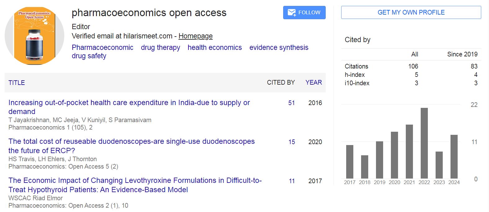
Pharmacoeconomics: Open Access received 106 citations as per Google Scholar report