Commentary - (2024) Volume 14, Issue 4
Received: 12-Sep-2024, Manuscript No. JBL-24-147889;
Editor assigned: 16-Sep-2024, Pre QC No. JBL-24-147889 (PQ);
Reviewed: 30-Sep-2024, QC No. JBL-24-147889;
Revised: 07-Oct-2024, Manuscript No. JBL-24-147889 (R);
Published:
25-Oct-2024
, DOI: 10.37421/2165-7831.2024.14.331
Citation: Ikegame Kazuhiro. "Which Human Leukocyte Antigen (HLA) are Donor-Derived T-cells Restricted to After HLA-Mismatched Hematopoietic Stem Cell Transplantation? " J Blood Lymph 14(2024): 331
Copyright: © 2024 Ikegame K. This is an open-access article distributed under the terms of the creative commons attribution license which permits unrestricted use, distribution and reproduction in any medium, provided the original author and source are credited.
With advances in preconditioning regimens and supportive care, transplants from related donors who share only one of the two Human Leukocyte Antigen (HLA) haplotypes known as HLA. Haploidentical Hematopoietic Stem Cell Transplantation (haplo-HCT) have been widely performed. Three HLA haplotypes are involved in haplo-HCT: The shared haplotype of the patient and donor (shared HLA), the haplotype uniquely possessed by the donor (donor-specific HLA) and the haplotype belonging to the patient (host-specific HLA). In this context, a critical question arises: Which HLA are donor-derived T cells restricted to after transplantation? Immediately following transplantation, mature donor T and stem cells are transferred to the recipient’s body. Approximately half of the donor-derived mature Tcell population is assumed to share the same HLA restriction, while the other half is expected to exhibit donor-specific HLA restriction. Shared HLA-restricted donor T cells act as a biological defense against pathogens in both the patient’s and donor’s bodies. Donorspecific HLA-restricted T cells can only recognize donor-derived hematopoietic cells in the patient’s body. If a hematotropic virus, such as Cytomegalovirus (CMV), infects donor blood cells after transplantation, donor-specific HLA-restricted T cells can eliminate the infected cells (Figure 1A). However, when an epitheliotropic virus, such as coronavirus, infects the epithelial cells of the recipient, viral peptides are present in both the shared HLA and host-specific HLA. Donor-specific HLA-restricted T cells fail to recognize the infection in an antigen-specific manner. Furthermore, donor-specific HLArestricted T cells that initially recognized an irrelevant antigen in the donor’s body may also identify host-specific HLA/self-peptide complexes (Figure 1B). The frequency of cross-reactivity is unexpectedly high (>1%) in donor T cell populations [1]. Such T cells may induce Graft-Versus-Host Disease (GVHD) and Graft- Versus-Leukemia (GVL) in a promiscuous manner [2,3].

Figure 1. Schematic presentation of a case with HLA completely mismatched transplantation and a speculated mechanism of alloreactive immunity (modified with [5]). Note: Recipient HLA: A*11:01/33:03, B*40:01/44:03, C*07:02/14:03, DRB1*09:01/13:02; Donor HLA: A*02:01/24:02, B*15:07/54:01, C*01:02/03:03, DRB1*04:03/15:01.
In contrast, host-specific HLA-restricted T cells eliminate the patient’s epithelial cells infected with an epitheliotropic virus (Figure 1C). Immediately after transplantation, donor-specific HLA-restricted T cells should not be present in the recipient’s body. However, precursor T cells differentiate from donor stem cells over time following transplantation. These precursor T cells enter the thymus for maturation. T Cell Receptor (TCR) reconstitution occurs randomly, with positive and negative selection of T cells with moderate affinity for the HLAs expressed in the thymus (shared HLA and host-specific HLA). If the HLAs expressed by the T cells themselves are not involved in this process, host-specific HLA-restricted T cells of donor origin may emerge. This phenomenon has been proven in mice [4] but not in humans, these are all shown in Figure 1.
Experimental results
Based on this background, we aimed to determine the proportions of shared HLA, donor-specific HLA and host-specific HLA-restricted T cells in post-transplant patients [5]. Flow cytometry with CMV-specific HLA-A*24:02 and HLA-A*02:01 tetramers was employed to examine peripheral blood samples over time in patients undergoing HLAmismatched transplantation, where patient-donor combinations correspond to shared HLAs, donor-specific HLAs, or host-specific HLAs. As expected, shared HLA and donor-specific HLA-restricted T cells were detected early after transplantation, while host-specific HLA-restricted T cells were not detected.
Transplantation beyond HLA haploidentical settings
Another focus of our study was to gather information on transplants beyond the requirement for half-matching HLA; specifically, transplants from relatives with differing HLA haplotypes (2-haplo-mis- HCT) and spousal transplants. Although these transplants may be unconventional, cases have been reported where the transplant was performed by mistake [6], or identified in retrospective studies [7]. We independently conducted a prospective phase I/II clinical trial of 2- haplo-mis-HCT and reported its safety and efficacy in 30 patients [8]. Currently, a phase II trial is in progress (UMIN000043973). We noted five cases [9] of spousal transplants and are conducting a phase I/II clinical trial (UMIN000023235). In our experience, even if HLAs differ between patients and donors, engraftment and GVHD are not problematic. However, immune reconstitution may be required after transplantation. As mentioned earlier, in situations where all HLAs are different meaning there is no shared HLA, if an epitheliotropic virus infects the patient’s epithelial cells, the presence of hostspecific HLA-restricted T cells is necessary for elimination.
In addition to addressing HLA-mismatched thymic selection in humans, we aimed to determine whether host-specific HLA-restricted T cells were present. Notably, we presented a case of COVID-19 following transplantation from a donor with a different HLA (Figure 1). The recipient developed COVID-19 and recovered without complications. The viral peptide should have been present in the host HLA during infection. Although we could not detect host-specific HLArestricted T cells in vitro, their presence was suggested in vivo.
Detection of antigen-specific T cells restricted to desired HLA
It is well known that the frequency of antigen-specific T cells detected by flow cytometry using tetramers is typically a small percentage of all CD3+ or CD8+ cells [10]. However, considerable evidence indicates that antigen-specific T cells remain active even at lower levels [11-13]. In recent COVID-19 studies, reactive T cells have been quantified using virus-specific TCR sequences [14]. Although the current flow cytometry-based study did not fulfill our objective of detecting host-specific HLA-restricted T cells, more sophisticated methods may be able to detect their presence. Since 2-haplo-mis-HCT and spousal transplants are last-resort options for patients who have exhausted curative treatment avenues due to a lack of suitable donors, theoretical advancements should be further pursued.
The patient underwent 2-HLA haplotype-mismatched Hematopoietic Stem Cell Transplantation (2-haplo-mis-HCT) from sibling with an incompatible HLA, as shown at the below of the figure.
• Donor-derived Dendritic Cells (DC, orange) present a Cytomegalovirus (CMV) peptide antigen with donor-specific HLA, A*24:02 (red HLA/dark purple peptide), which activates donorspecific HLA-restricted T cells (T24 purple) to attack CMV-infected donor blood cells.
• Donor T cells (T2 gray), which originally recognize the A*02:01/ irrelevant peptide complex (green HLA/pink peptide), might promiscuously (from the side chain of the HLA/peptide complex) cross-react with the A*11:01/self-peptide (blue HLA/black peptide). These cells may function as GVHD and GVL clones.
• Precursor T cells derived from donor stem cells (preT green) enter the recipient thymus and are educated to be HLA (A*33:03 or A*11:01) restricted (T11 sky blue). Donor DC take up the circulating recipient HLA-A*11:01 to become hybrid DC, presenting antigens with both donor and recipient HLAs [15]. The host-specific HLA-restricted T cells (T11 sky blue) attack the epithelial cells of virus-infected recipients.
[Crossref] [Google Scholar] [Pubmed]
[Crossref] [Google Scholar] [Pubmed]
[Crossref] [Google Scholar] [Pubmed]
[Crossref] [Google Scholar] [Pubmed]
[Crossref] [Google Scholar] [Pubmed]
[Crossref] [Google Scholar] [Pubmed]
[Crossref] [Google Scholar] [Pubmed]
[Crossref] [Google Scholar] [Pubmed]
[Crossref] [Google Scholar] [Pubmed]
[Crossref] [Google Scholar] [Pubmed]
[Crossref] [Google Scholar] [Pubmed]
[Crossref] [Google Scholar] [Pubmed]
[Crossref] [Google Scholar] [Pubmed]
[Crossref] [Google Scholar] [Pubmed]
[Crossref] [Google Scholar] [Pubmed]
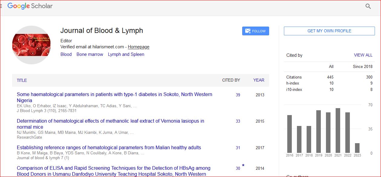
Journal of Blood & Lymph received 443 citations as per Google Scholar report