Basant kumar Sinha
Bihar Veterinary College, India
Posters & Accepted Abstracts: J Med Microb Diagn
Aflatoxicosis was produced in apparently healthy murrah buffalo male calves weighing about 30-40 kg, by administering AFB1 orally as 10 microgram/kg of body weight daily in glucopropylene base till the end of the experiment i.e 30 days or animal died. Control animals received only glycopropylene in the same quantity. Immunological profile (both humoral and cell mediated immune response) was studied in experimentally produced animal at different interval. Humoral response was judged by EAC-rosette and antibody titre against SRBC, CMIR was judged by E-rosette and DTH at 0,10,20 and 30 days intoxication. EAC-rosette was 61-33 �?± 1.77, 59.66 �?± 0.88, 54.33 �?± 2.37 and 42.33 �?± 4.10 respectively. The level was found to be statistically significant (p <0.05) only after 30 days of post infection. HA titre Log2/0.05 in control buffalo after 16 days (1st injection) and 25 days (2nd injection) was 8.33 �?± 0.33 and 9.66 �?± 0.33 respectively, whereas corresponding HA titre in aflatoxin fed calves was 7.33 �?± 0.33 and 6.33 �?± 0.33 respectively. The titre was found to be statistically significant (p <0.005) after end dose of SRBC administration. Per cent E-rosette at 0,10,20 and 30 days post intoxication was 18.21 �?± 3.16 which increased upto 10 days i.e. 23.73 �?± 2.97, but in later stage of DPI i.e at 30 and 30 days it became 15.2 �?± 1.43 and 10.63 �?± 0.89 respectively. It is evident that during early stage of post-intoxication, T-cell population increased, this may be due to inflammatory response set in as a result of infection but in later stage the decrease in per cent E-rosette explains the suppressive effect of AFB1 on CMIR and these responses were dose dependent. DTH study revealed skin thickness before and after sensitization in control buff as 3.56 �?± 0.35 and 10.5 �?± 0.75 respectively, whereas in aflatoxin fed animals the corresponding value was 3.43 �?± 0.12 and 4.56 �?±0.32 respectively. The above studies clearly revealed immunosuppression by aflatoxin. In another study in rabbit, impairment of Reticuloendothelial System (RES) was also recorded. Biochemical profile in experimentally induced calves revealed significant increase of alkaline phosphatase, alanine amino transferase (SGPT) and aspartate amino transferase (SGOT) after 20 and 30 days of post feeding of aflatoxin. Decreases albumin and increased globulin level resulted into complete reversion of A:G ratio. This indicates the impairment of liver during aflatoxicosis. Histopathological studies of different organ revealed that among all the organs liver was the main target of action and maximum histopathological changes i.e. cirrhosis to neoplastic changes was detected in liver. Lungs showed pneumonic changes. Kidney showed tubular degeneration and necrosis of lining of epithelial tubules. Germinal centers of spleen was completely lost and there was little changes in stomach and intestine Meninges of the brain showed slight congestion.
Email: bksmicro@gmail.com
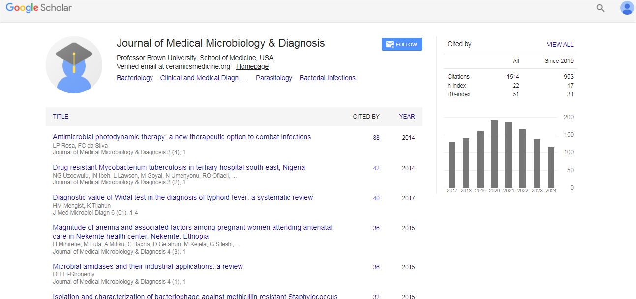
Medical Microbiology & Diagnosis received 14 citations as per Google Scholar report

 Spanish
  Chinese
  Russian
  German
  French
  Japanese
  Portuguese
  Hindi
Hindi 