Dalip Singh Mehta
Indian Institute of Technology Delhi, India
Keynote: J Laser Opt Photonics
Bright field optical microscopy has played significant role in biological research. But this technique provides qualitative information about the biological samples, such as shape, morphology, etc. Most of the biological cells and tissues are transparent in nature, i.e., they do not absorb the amplitude of light significantly, therefore, the fine details of cells and tissues cannot be visualized using bright field microscopy because of the poor contrast. To visualize such structures Zernike developed Phase Contrast Microscopy (PCM). In PCM the image contrast can be improved by means of converting spatial phase shift of light field into an interference pattern, thus fine structure of the cells and tissues can obtained without using any exogenous contrast. But this technique also gives only qualitative information about the cells and tissues. Recently, Quantitative Phase Microscopy (QPM) and nanoscopy has greatly contributed for the measurement of various parameters of biological cells and tissues quantitatively for early stage disease detection. In this presentation various QPM techniques, such as, digital holographic microscopy, white light interference microscopy, spatially low coherent light interference microscopy, diffraction phase microscopy and QPM combined with evanescent field trapped red blood cells and Total Internal Reflection Fluorescence (TIRF) microscopy will be reviewed and their applications in biological research will be presented. More recently, structured illumination microscopy (also called nanoscopy) combined with digital holographic microscopy is being investigated for quantitative phase nanoscopy of biological cells and tissues. Some of these techniques and their importance will be presented. Finally the summary of all these techniques and their future prospects will be reviewed. Recent Publications 1. Azeem Ahmad, Vishal Srivastava, Vishesh Dubey and D S Mehta (2015) Ultra-short longitudinal spatial coherence length of laser light with the combined effect of spatial, angular, and temporal diversity. Applied Physics Letters; 106, 093701. 2. D S Mehta and Vishal Srivastava (2012) Quantitative phase imaging of human red blood cells using phase-shifting white light interference microscopy with colour fringe analysis. Applied Physics Letters; 101: 203701.
Dalip Singh Mehta is currently a Professor at the Department of Physics, Indian Institute of Technology Delhi, India. Previously, he has worked as an Associate Professor and Assistant Professor at Indian Institute of Technology Delhi. He has contributed more than 110 research papers in international refereed journals and more than 150 in international and national conferences. He has delivered more than 45 invited talks/lectures in various international and national conferences and universities. He has received Teaching Excellence Award 2013 from the Indian Institute of Technology Delhi, India. Email:mehtads@physics.iitd.ac.in
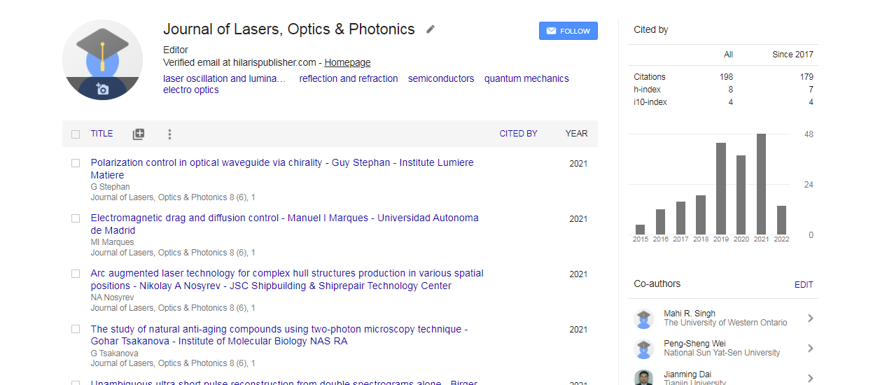
Journal of Lasers, Optics & Photonics received 279 citations as per Google Scholar report

 Spanish
  Chinese
  Russian
  German
  French
  Japanese
  Portuguese
  Hindi
Hindi 